 뒤로
뒤로Cell Structure, Function, and Microscopy: General Biology Study Guide
스터디 가이드 - 스마트 노트
 자료에 맞춘 맞춤형 노트, 핵심 정의, 예시, 맥락을 확장해 제공합니다.
Q1. Describe an overview of key cell structures that are necessary to perform life functions.
Background
Topic: Cell Structure and Function
This question tests your understanding of the essential components of cells and their roles in maintaining life processes.
Key Terms:
Plasma membrane
Cytoplasm
Ribosomes B
DNA
Step-by-Step Guidance
Identify the basic structures found in all cells: plasma membrane, cytoplasm, ribosomes, and DNA.
Explain the function of each structure: for example, the plasma membrane regulates entry and exit of substances, ribosomes synthesize proteins, etc.
Consider why each structure is essential for cell survival and function.
Think about how these structures i nteract to support cellular processes.
Try solving on your own before revealing the answer!
Final Answer:
All cells have a plasma membrane (controls what enters/exits), cytoplasm (site for cellular processes), ribosomes (make proteins), and DNA (genetic information). These structures are essential for life because they allow cells to maintain homeostasis, produce proteins, and pass on genetic information.
Q2. Using the table, give the specific cell size ranges for the following and rank them smallest to largest: atom, ribosomes, small bacteria, mitochondria, animal cells, chicken egg.
Background
Topic: Cell Size and Scale
This question tests your ability to compare the sizes of biological structures and understand their relative scale.
Key Terms:
Atom: smallest unit of matter
Ribosome: protein synthesis machinery
Bacteria: unicellular organisms
Mitochondria: organelle for energy production
Animal cell: eukaryotic cell
Chicken egg: large single cell
Step-by-Step Guidance
Look up or recall the approximate size ranges for each structure (e.g., atom ~0.1 nm, ribosome ~20 nm, etc.).
Arrange the structures in order from smallest to largest based on their size.
Check your ranking against the typical biological scale.
Consider why size matters for visualization and function.
Try solving on your own before revealing the answer!
Final Answer:
Smallest to largest: atom < ribosome < small bacteria < mitochondria < animal cell < chicken egg. Atoms are the smallest, while chicken eggs are the largest single cells.
Q3. Describe the difference between the different microscopes listed below. Give examples of the structures that are visible by each type of microscope and the rationale for their use.
Background
Topic: Microscopy and Cell Visualization
This question tests your understanding of how different microscopes are used to study cell structures.
Key Terms:
Light microscope
Fluorescence microscope
Electron microscope (SEM, TEM)
Step-by-Step Guidance
Define each type of microscope and its principle of operation.
List the cell structures that can be visualized with each microscope (e.g., light microscopes for cells, electron microscopes for organelles).
Explain why certain microscopes are chosen for specific structures (resolution, detail, live/dead cells).
Consider examples from textbook figures or images.
Try solving on your own before revealing the answer!
Final Answer:
Light microscopes are used for whole cells; fluorescence microscopes highlight specific molecules; electron microscopes (SEM for surface, TEM for internal structures) are used for high-resolution images of organelles and small structures.
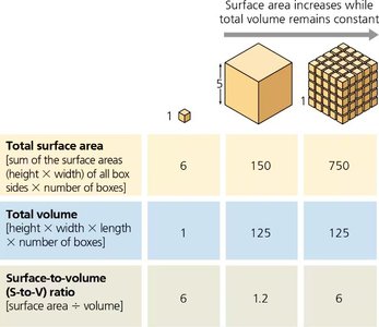
Q8. Why must cells be small? Why not have one big cell to do the job for the entire body?
Background
Topic: Cell Size and Surface Area-to-Volume Ratio
This question tests your understanding of why cells are small and how their size affects function.
Key Terms and Formula:
Surface area-to-volume ratio
Diffusion
Step-by-Step Guidance
Recall that cells rely on diffusion to move materials in and out.
Understand that as cells get larger, their volume increases faster than their surface area.
Calculate surface area and volume for different cell sizes using the formulas above.
Compare the ratios for small and large cells to see how efficiency changes.
Think about the consequences for cellular function if the ratio is too low.
Try solving on your own before revealing the answer!
Final Answer:
Cells must be small because a high surface area-to-volume ratio allows efficient transport of materials. Large cells would not be able to move materials quickly enough to support life processes.
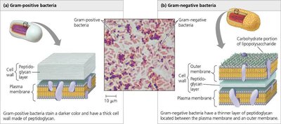
Q17. Using the figure, describe the structural differences in gram positive vs gram negative bacteria.
Background
Topic: Bacterial Cell Walls and Antibiotic Action
This question tests your understanding of bacterial cell wall structure and its relevance to antibiotic effectiveness.
Key Terms:
Gram-positive bacteria
Gram-negative bacteria
Peptidoglycan
Outer membrane
Step-by-Step Guidance
Examine the diagram showing the cell wall structures of gram-positive and gram-negative bacteria.
Identify the thickness of the peptidoglycan layer in each type.
Note the presence or absence of an outer membrane.
Consider how these differences affect staining and antibiotic susceptibility.
Try solving on your own before revealing the answer!
Final Answer:
Gram-positive bacteria have a thick peptidoglycan layer and no outer membrane; gram-negative bacteria have a thin peptidoglycan layer and an additional outer membrane. This affects how antibiotics like penicillin work.
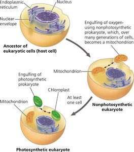
Q25. Describe the endosymbiosis theory and how the current eukaryotic cell may have derived with early origins with a prokaryotic cell. Use the diagram in your description.
Background
Topic: Evolution and Endosymbiosis Theory
This question tests your understanding of how eukaryotic cells evolved from prokaryotic ancestors.
Key Terms:
Endosymbiosis
Mitochondria
Chloroplast
Prokaryote
Eukaryote
Step-by-Step Guidance
Review the diagram showing the engulfing of prokaryotic cells by a host cell.
Describe how mitochondria and chloroplasts originated from prokaryotes living inside another cell.
Explain the evidence supporting this theory (e.g., double membranes, DNA similarities).
Connect the theory to modern eukaryotic cell structure.
Try solving on your own before revealing the answer!
Final Answer:
The endosymbiosis theory proposes that mitochondria and chloroplasts originated from prokaryotic cells engulfed by a host cell. Evidence includes their own DNA, double membranes, and similarities to prokaryotes.
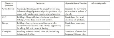
Q50. What organelles or cell structures are likely malfunctioning in these diseases (Cystic Fibrosis, ALD, Pompe, Kartagener)?
Background
Topic: Cell Structure and Disease
This question tests your ability to connect symptoms and disease to malfunctioning organelles.
Key Terms:
Cystic Fibrosis: plasma membrane
ALD: peroxisomes
Pompe: lysosomes
Kartagener: cilia (cytoskeleton)
Step-by-Step Guidance
Read the symptoms and normal function for each disease.
Match the symptoms to the organelle responsible (e.g., movement, breakdown of molecules).
Consider how malfunction of each organelle leads to the observed symptoms.
Fill in the affected organelle for each disease.
Try solving on your own before revealing the answer!
Final Answer:
Cystic Fibrosis: plasma membrane; ALD: peroxisomes; Pompe: lysosomes; Kartagener: cilia/cytoskeleton. Each disease is linked to malfunction of a specific organelle.
