 뒤로
뒤로The Cell Cycle and Mitosis: Mechanisms of Cellular Continuity
스터디 가이드 - 스마트 노트
 자료에 맞춘 맞춤형 노트, 핵심 정의, 예시, 맥락을 확장해 제공합니다.
Chapter 12: The Cell Cycle
Overview of the Cell Cycle
The cell cycle is the series of events that cells go through as they grow and divide. It ensures the accurate transmission of genetic information from one generation of cells to the next, supporting growth, development, and tissue repair in multicellular organisms.
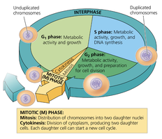
Interphase: The cell grows, performs its normal functions, and duplicates its DNA.
M phase (Mitotic phase): The cell divides its nucleus (mitosis) and cytoplasm (cytokinesis), producing two genetically identical daughter cells.
Concept 12.1: Mitosis and Genetic Identity
Chromosomes, Chromatids, and Chromatin
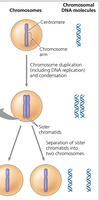
Genetic information is organized into chromosomes, which are duplicated and distributed during cell division. Understanding the terminology is essential for grasping how mitosis produces genetically identical cells.
Chromosome: A tightly coiled structure of DNA and proteins that carries genetic information.
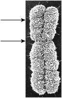
Chromatid: One of two identical halves of a duplicated chromosome, joined at the centromere.
Chromatin: The loose, uncoiled form of DNA present in the nucleus during interphase.
Centromere: The region where sister chromatids are joined together.
Key Roles of Cell Division
Asexual reproduction: Single-celled organisms reproduce by dividing (e.g., Amoeba).
Growth and development: Multicellular organisms grow by increasing cell number (e.g., embryo development).
Tissue renewal and repair: Replacement of dead or damaged cells (e.g., skin healing).
Genome and Chromosome Number
Genome: The complete set of genetic material in an organism.
Somatic cells: Body cells with two sets of chromosomes (diploid); humans have 46 chromosomes per somatic cell.
Gametes: Sex cells (sperm and egg) with one set of chromosomes (haploid); humans have 23 chromosomes per gamete.
Distinguishing Terms
Chromosome: One DNA molecule, tightly coiled.
Duplicated chromosome: Two sister chromatids joined at the centromere.
After separation: Each chromatid is considered an individual chromosome.
Mitosis vs. Cytokinesis
Mitosis: Division of the nucleus and its genetic material.
Cytokinesis: Division of the cytoplasm, resulting in two separate cells.
Meiosis
Meiosis is a specialized form of cell division that produces gametes with half the chromosome number, occurring in the ovaries and testes in humans.
Concept 12.2: Phases of the Cell Cycle
Phases of the Cell Cycle
The cell cycle consists of interphase (G1, S, G2) and the M phase (mitosis and cytokinesis).
Phase | Key Events |
|---|---|
G1 | Cell growth, normal functions, protein and organelle synthesis |
S | DNA replication, chromosome duplication |
G2 | Further growth, preparation for mitosis, error checking |
M | Mitosis (nuclear division) and cytokinesis (cytoplasmic division) |
Mitotic Spindle and Centrosomes
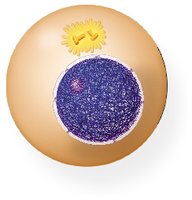
The mitotic spindle is a structure made of microtubules that segregates chromosomes during mitosis. Centrosomes, also known as microtubule-organizing centers, play a key role in spindle formation and chromosome movement.
Phases of Mitosis
Mitosis is divided into several phases, each with distinct events:
Phase | Key Features |
|---|---|
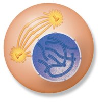
Prophase | Chromatin condenses, nucleoli disappear, spindle forms, centrosomes move apart |
Prometaphase | Nuclear envelope breaks down, chromosomes condense further, kinetochores form, spindle fibers attach |
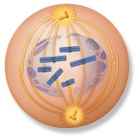
Metaphase | Chromosomes align at the metaphase plate, spindle fully formed |
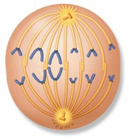
Anaphase | Sister chromatids separate and move to opposite poles |
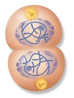
Telophase | Nuclear envelopes reform, chromosomes decondense, nucleoli reappear |
Cytokinesis | Cytoplasm divides, forming two daughter cells |
Kinetochore vs. Nonkinetochore Microtubules
Kinetochore microtubules: Attach to chromosomes at the kinetochore and pull sister chromatids apart.
Nonkinetochore microtubules: Overlap at the cell center and help elongate the cell during division.
Cytokinesis in Animal and Plant Cells
Animal cells: Cleavage furrow forms, pinching the cell in two.
Plant cells: Cell plate forms from Golgi-derived vesicles, developing into a new cell wall.
Binary Fission in Prokaryotes
Prokaryotes divide by binary fission, a simpler process involving DNA replication and division into two genetically similar cells.
Concept 12.3: Regulation of the Cell Cycle
Cell Cycle Checkpoints
The cell cycle is regulated by checkpoints that ensure each phase is completed correctly before the next begins. The main checkpoints are:
Checkpoint | Function |
|---|---|
G1 | Checks cell size, nutrients, and DNA integrity |
G2 | Checks DNA replication accuracy |
M | Checks chromosome attachment to spindle fibers |
Molecular Control: Cyclins and Cdks
Progression through the cell cycle is controlled by cyclins and cyclin-dependent kinases (Cdks). Cyclins accumulate and bind to Cdks, forming active complexes (e.g., MPF) that trigger cell cycle events such as mitosis. Cyclin levels fluctuate, causing Cdk activity to rise and fall.
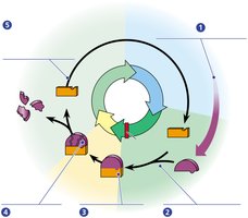
Protein kinases: Enzymes that activate other proteins by phosphorylation.
MPF (Maturation Promoting Factor): Triggers mitosis by phosphorylating target proteins.
G0 phase: A non-dividing state; some cells (e.g., neurons, muscle cells) remain here permanently.
Growth Factors and Cancer
Growth factors: Proteins that stimulate cell division (e.g., PDGF for fibroblasts).
Density-dependent inhibition: Normal cells stop dividing when crowded; cancer cells do not.
Anchorage dependence: Normal cells require attachment to a substrate to divide; cancer cells can divide without attachment.
Transformation: Conversion of a normal cell to a cancer cell.
Metastasis: Spread of cancer cells to other parts of the body.
Benign vs. malignant tumors: Benign tumors do not spread; malignant tumors invade other tissues.
Cancer treatments: Chemotherapy and radiation therapy damage DNA in rapidly dividing cells.
