 뒤로
뒤로The Cell: Structure, Function, and Evolutionary Origins
스터디 가이드 - 스마트 노트
 자료에 맞춘 맞춤형 노트, 핵심 정의, 예시, 맥락을 확장해 제공합니다.
The Cell: Structure, Function, and Evolutionary Origins
Overview of Cell Structure
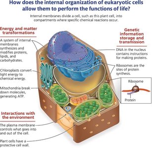
Cells are the fundamental units of life, performing all necessary functions to sustain living organisms. Both prokaryotic and eukaryotic cells share certain features, but also have distinct differences that reflect their evolutionary history and specialization.
DNA: Stores genetic information and directs cellular processes.
Ribosomes: Synthesize proteins essential for cell function.
Plasma Membrane: Regulates the entry and exit of materials, maintaining the internal environment.
Cytoplasm: The site where nutrients, organelles, and cellular processes are housed.
Visualizing Cells with Microscopes
Microscopes are essential tools for studying cells and their structures. Different types of microscopes provide varying levels of detail and are suited for specific cellular components.
Light Microscope: Used to view living cells, tissues, and larger organelles (e.g., nucleus, whole cells).
Fluorescence Microscope: Highlights specific structures using fluorescent markers, useful for tracking proteins or organelles.
Electron Microscope: Provides high-resolution images of small structures (e.g., ribosomes, membranes).
Scanning Electron Microscope (SEM): Visualizes cell surfaces in 3D detail.
Transmission Electron Microscope (TEM): Reveals internal structures at the molecular level.
Cell Size and Surface Area-to-Volume Ratio
Cell size is limited by the surface area-to-volume ratio, which affects the efficiency of material exchange and cellular communication. As cells grow, their volume increases faster than their surface area, reducing the efficiency of nutrient uptake and waste removal.
Surface Area: The total area available for exchange of materials.
Volume: The space inside the cell that must be serviced by the surface area.
Surface Area-to-Volume Ratio: Decreases as cell size increases, limiting cell growth.
Formula for Volume of a Cube:
Formula for Surface Area of a Cube:
Surface Area-to-Volume Ratio:
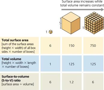
Key Point: As cell size increases, the surface area-to-volume ratio decreases, making it harder for the cell to efficiently exchange materials with its environment.
Prokaryotic Cells
Prokaryotes, including bacteria and archaea, are simpler cells lacking membrane-bound organelles. They are typically smaller than eukaryotic cells and have unique features that are targeted by antibiotics.
Cell Wall: Provides structure and protection; composed of peptidoglycan in bacteria.
Gram-Positive Bacteria: Thick peptidoglycan layer; stains darkly; more susceptible to antibiotics like penicillin.
Gram-Negative Bacteria: Thin peptidoglycan layer and an outer membrane; less susceptible to certain antibiotics.
Chromosomes: Circular DNA located in the nucleoid region.
Ribosomes: Smaller and structurally different from eukaryotic ribosomes.
Other Structures: Capsule (protection), flagella (movement), plasmids (extra DNA).
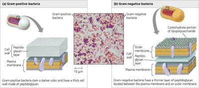
Antibiotic Action: Antibiotics exploit differences in cell wall structure and ribosomes to selectively target bacteria without harming human cells.
Eukaryotic Cells: Plant vs. Animal
Eukaryotic cells are more complex, containing membrane-bound organelles that compartmentalize cellular functions. Plant and animal cells share many organelles but also have distinct differences.
Shared Organelles: Nucleus, mitochondria, endoplasmic reticulum, Golgi apparatus, cytoskeleton, plasma membrane.
Plant-Specific: Chloroplasts (photosynthesis), cell wall (cellulose), large central vacuole.
Animal-Specific: Lysosomes, centrioles.
Membrane-Bound Organelles and Compartmentalization
Membrane-bound organelles allow eukaryotic cells to compartmentalize different biochemical processes, increasing efficiency and specialization.
Nucleus: Stores genetic material and coordinates cell activities.
Endoplasmic Reticulum (ER): Rough ER synthesizes proteins; smooth ER synthesizes lipids and detoxifies chemicals.
Golgi Apparatus: Modifies, sorts, and packages proteins for secretion.
Lysosomes: Digest cellular waste and foreign material.
Mitochondria: Generate ATP through cellular respiration.
Chloroplasts: Convert solar energy to chemical energy in plants.
The Cytoskeleton
The cytoskeleton is a network of protein filaments that provides structural support, enables movement, and organizes cell components.
Microtubules: Maintain cell shape, move organelles, and are components of cilia and flagella.
Actin Filaments (Microfilaments): Involved in cell movement, muscle contraction, and cell division.
Intermediate Filaments: Provide mechanical support and maintain cell integrity.
Consequences of Cytoskeleton Loss: Disrupted cell shape, impaired movement, and defective intracellular transport.
Endosymbiosis Theory and Evolution of Eukaryotes
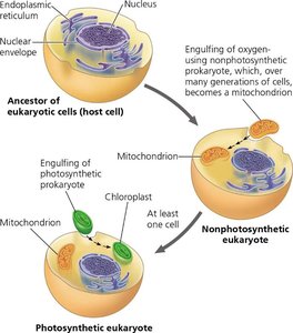
The endosymbiosis theory explains the origin of mitochondria and chloroplasts as formerly free-living prokaryotes that were engulfed by ancestral eukaryotic cells. This symbiotic relationship led to the evolution of complex eukaryotic cells.
Evidence for Endosymbiosis:
Mitochondria and chloroplasts have their own circular DNA, similar to bacteria.
They replicate independently of the cell via binary fission.
They have double membranes, consistent with engulfment.
Their ribosomes resemble those of prokaryotes.
Antibiotics and the Battle Against Bacteria
Antibiotics are drugs that target specific features of bacterial cells, such as cell wall synthesis, protein synthesis, or DNA replication. Because human cells lack these bacterial features, antibiotics can selectively kill bacteria without harming human cells.
Penicillin: Inhibits peptidoglycan synthesis in bacterial cell walls (effective against gram-positive bacteria).
Other Antibiotics: May target bacterial ribosomes or DNA replication enzymes.
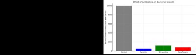
Natural Selection and Antibiotic Resistance: Bacteria can evolve resistance to antibiotics through natural selection, leading to the survival and proliferation of resistant strains.
Cellular Disorders and Organelle Malfunction
Diseases can result from malfunctioning organelles, affecting cell and tissue function. Understanding the role of each organelle helps diagnose and treat these conditions.
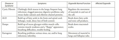
Disease or Syndrome | Symptoms | Organelle Normal Function | Affected Organelle |
|---|---|---|---|
Cystic Fibrosis | Thick mucus in lungs, digestive problems, salty sweat | Regulates movement of materials in and out of cell | Plasma membrane (channel proteins) |
ALD | Fatty acid buildup, lethargy, loss of limb control | Break down fatty acids and detox cell products | Peroxisomes |
Pompé | Glycogen buildup, muscle weakness, breathing difficulties | Break down macromolecules | Lysosomes |
Kartagener | Breathing problems, infertility | Movement of material in lungs and fallopian tubes | Cilia (microtubules) |
Application: Case Study Example
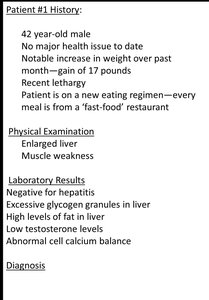
Patient #1 presents with excessive glycogen granules and high fat in the liver, muscle weakness, and abnormal calcium balance. These symptoms suggest a malfunction in organelles responsible for metabolism and detoxification, such as the smooth endoplasmic reticulum (SER) and possibly mitochondria.
Summary Table: Prokaryotes vs. Eukaryotes
Feature | Prokaryote | Eukaryote |
|---|---|---|
DNA | Circular, small | Linear, large |
Ribosomes | Smaller, different structure | Larger |
Size | Smaller, similar to mitochondria | Larger |
Cell Wall | Peptidoglycan (Gram +), outer membrane (Gram -) | Cellulose (plants), none (animals) |
Organelles | No membrane-bound organelles | Membrane-bound organelles present |
Key Takeaways
Cells are small to maximize surface area-to-volume ratio for efficient exchange of materials.
Microscopes are essential for visualizing cell structures at different scales.
Prokaryotic and eukaryotic cells differ in complexity, organelles, and genetic organization.
Antibiotics target unique features of bacterial cells, sparing human cells.
Organelle malfunction can lead to specific diseases, highlighting the importance of cellular compartmentalization.
The endosymbiosis theory explains the evolutionary origin of mitochondria and chloroplasts in eukaryotic cells.
