Ch. 10 - Eukaryotic Chromosome Abnormalities and Molecular Organization
Sanders - Genetic Analysis: An Integrated Approach 3rd Edition
Sanders3rd EditionGenetic Analysis: An Integrated ApproachISBN: 9780135564172당신이 사용하는 게 아니라요?교과서 변경
10장, 문제 26b

DNase I cuts DNA that is not protected by bound proteins but is unable to cut DNA that is complexed with proteins. Human DNA is isolated, stripped of its nonhistone proteins, and mixed with DNase I. Samples are removed after 30 minutes, 1 hour, and 4 hours and run separately in gel electrophoresis. The resulting gel is stained to make all DNA fragments in it visible, and the results are shown in the figure. DNA fragment sizes in base pairs (bp) are estimated by the scale to the left of the gel. Draw a conclusion about the organization of chromatin in the human genome from this gel.

검증된 단계별 안내
1
Step 1: Understand the role of DNase I in the experiment. DNase I is an enzyme that cuts DNA at regions that are not protected by proteins, such as histones. This means that regions of DNA tightly bound to histones or other proteins will remain intact, while unprotected regions will be fragmented.
Step 2: Analyze the experimental setup. Human DNA is stripped of nonhistone proteins, leaving primarily histones bound to the DNA. The DNA is then treated with DNase I at different time intervals (30 minutes, 1 hour, and 4 hours). The resulting DNA fragments are separated by gel electrophoresis, which sorts fragments by size, and the gel is stained to visualize the fragments.
Step 3: Interpret the gel electrophoresis results. Larger DNA fragments indicate regions of DNA that are protected by histones and not cut by DNase I, while smaller fragments represent regions of DNA that were accessible to DNase I and were cut. Over time, the presence of smaller fragments suggests that DNase I progressively cuts accessible regions of DNA.
Step 4: Relate the results to chromatin organization. The presence of both large and small DNA fragments suggests that human chromatin is organized into regions of tightly bound DNA (protected by histones) and regions of more accessible DNA. This supports the idea that chromatin is structured into nucleosomes, where DNA is wrapped around histones, interspersed with linker DNA that is more accessible.
Step 5: Draw a conclusion. The gel results demonstrate that human chromatin is not uniformly protected by histones. Instead, it is organized into a dynamic structure with regions of tightly bound DNA (nucleosomes) and regions of accessible DNA (linker DNA), which reflects the functional organization of the genome for processes like transcription and replication.

비슷한 문제에 대한 검증된 영상 답변:

이 영상 해법은 위 문제에 도움이 된다고 튜터들이 추천한 것입니다.
영상 길이:
1m
도움이 되었나요?

주요 개념

질문에 올바르게 답하기 위해 반드시 이해해야 하는 핵심 개념들은 다음과 같습니다.

Chromatin Structure

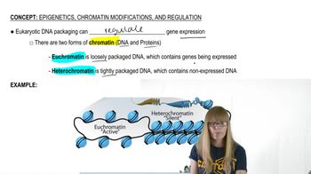
Chromatin is a complex of DNA and proteins found in the nucleus of eukaryotic cells. It exists in two forms: euchromatin, which is loosely packed and accessible for transcription, and heterochromatin, which is tightly packed and generally inactive. The organization of chromatin influences gene expression and DNA accessibility, making it crucial for understanding how DNase I interacts with DNA.
추천 영상:

DNase I Activity

DNase I is an enzyme that cleaves DNA, specifically targeting regions that are not protected by proteins. Its activity is indicative of the accessibility of DNA within chromatin. When DNA is complexed with proteins, such as histones, DNase I cannot cut it, which helps researchers infer the structural organization of chromatin based on the presence or absence of DNA fragments in gel electrophoresis.
추천 영상:
가이드 코스
08:04
Decision Between Lytic and Lysogenic Cycles

Gel Electrophoresis

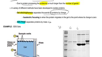
Gel electrophoresis is a laboratory technique used to separate DNA fragments based on their size. When an electric current is applied, smaller fragments move faster through the gel matrix than larger ones. By analyzing the pattern of DNA bands after staining, researchers can determine the sizes of the fragments and draw conclusions about the chromatin structure and the extent of DNase I digestion over time.
추천 영상:
관련 실천
교과서 질문

Genomic DNA from the nematode worm Caenorhabditis elegans is organized by nucleosomes in the manner typical of eukaryotic genomes, with 145 bp encircling each nucleosome and approximately 55 bp in linker DNA. When C. elegans chromatin is carefully isolated, stripped of nonhistone proteins, and placed in an appropriate buffer, the chromatin decondenses to the 10-nm fiber structure. Suppose researchers mix a sample of 10-nm–fiber chromatin with a large amount of the enzyme DNase I that randomly cleaves DNA in regions not protected by bound protein. Next, they remove the nucleosomes, separate the DNA fragments by gel electrophoresis, and stain all the DNA fragments in the gel.

Approximately what range of DNA fragment sizes do you expect to see in the stained electrophoresis gel? How many bands will be visible on the gel?

465
views
교과서 질문

A boy with Down syndrome (trisomy 21) has 46 chromosomes. His parents and his two older sisters have a normal phenotype, but each has 45 chromosomes.

What is the probability the next child of this couple will have a normal phenotype and have 46 chromosomes? Explain your answer.

513
views
교과서 질문

Genomic DNA from the nematode worm Caenorhabditis elegans is organized by nucleosomes in the manner typical of eukaryotic genomes, with 145 bp encircling each nucleosome and approximately 55 bp in linker DNA. When C. elegans chromatin is carefully isolated, stripped of nonhistone proteins, and placed in an appropriate buffer, the chromatin decondenses to the 10-nm fiber structure. Suppose researchers mix a sample of 10-nm–fiber chromatin with a large amount of the enzyme DNase I that randomly cleaves DNA in regions not protected by bound protein. Next, they remove the nucleosomes, separate the DNA fragments by gel electrophoresis, and stain all the DNA fragments in the gel.

How do the expected results support the 10-nm–fiber model of chromatin?

457
views
교과서 질문

Genomic DNA from the nematode worm Caenorhabditis elegans is organized by nucleosomes in the manner typical of eukaryotic genomes, with 145 bp encircling each nucleosome and approximately 55 bp in linker DNA. When C. elegans chromatin is carefully isolated, stripped of nonhistone proteins, and placed in an appropriate buffer, the chromatin decondenses to the 10-nm fiber structure. Suppose researchers mix a sample of 10-nm–fiber chromatin with a large amount of the enzyme DNase I that randomly cleaves DNA in regions not protected by bound protein. Next, they remove the nucleosomes, separate the DNA fragments by gel electrophoresis, and stain all the DNA fragments in the gel.

Explain the origin of DNA fragments seen in the gel.

596
views
교과서 질문

Experimental evidence demonstrates that the nucleosomes present in a cell after the completion of S phase are composed of some 'old' histone dimers and some newly synthesized histone dimers. Describe the general design for an experiment that uses a protein label such as ³⁵S to show that nucleosomes are often a mixture of old and new histone dimers following DNA replication.

501
views
교과서 질문

DNase I cuts DNA that is not protected by bound proteins but is unable to cut DNA that is complexed with proteins. Human DNA is isolated, stripped of its nonhistone proteins, and mixed with DNase I. Samples are removed after 30 minutes, 1 hour, and 4 hours and run separately in gel electrophoresis. The resulting gel is stained to make all DNA fragments in it visible, and the results are shown in the figure. DNA fragment sizes in base pairs (bp) are estimated by the scale to the left of the gel. Examine the gel results and speculate why longer DNase I treatment produces different results.

443
views