
Duchenne muscular dystrophy (DMD; OMIM 310200) and Becker muscular dystrophy (BMD; OMIM 300376) are both X-linked recessive conditions that result from different mutations of the same gene, known as dystrophin, on the long arm of the chromosome. BMD and DMD are quite different clinically. DMD is a very severe disorder that first appears at a young age, progresses rapidly, and is often fatal in the late teens to 20s. BMD, on the other hand, is much milder. Often symptoms don't first appear until the 40s or 50s, the progression of the disease is slow, and fatalities due to BMD are infrequent. Go to https://www.ncbi.nlm.nih/omim and survey the information describing the gene mutations causing these two conditions. Discuss the information you find with a few others in a small group, and write a single summary explaining your findings.

- Ch. 1 - The Molecular Basis of Heredity, Variation, and Evolution64
- Ch. 2 - Transmission Genetics144
- Ch. 3 - Cell Division and Chromosome Heredity81
- Ch. 4 - Gene Interaction87
- Ch. 5 - Genetic Linkage and Mapping in Eukaryotes103
- Ch. 6 - Genetic Analysis and Mapping in Bacteria and Bacteriophages73
- Ch. 7 - DNA Structure and Replication71
- Ch. 8 - Molecular Biology of Transcription and RNA Processing79
- Ch. 9 - The Molecular Biology of Translation110
- Ch. 10 - Eukaryotic Chromosome Abnormalities and Molecular Organization100
- Ch. 11 - Gene Mutation, DNA Repair, and Homologous Recombination86
- Ch. 12 - Regulation of Gene Expression in Bacteria and Bacteriophage90
- Ch. 13 - Regulation of Gene Expression in Eukaryotes38
- Ch. 14 - Analysis of Gene Function via Forward Genetics and Reverse Genetics72
- Ch. 15 - Recombinant DNA Technology and Its Applications69
- Ch. 16 - Genomics: Genetics from a Whole-Genome Perspective49
- Ch. 17 - Organelle Inheritance and the Evolution of Organelle Genomes27
- Ch. 18 - Developmental Genetics43
- Ch. 19 - Genetic Analysis of Quantitative Traits66
- Ch. 20 - Population Genetics and Evolution at the Population, Species, and Molecular Levels105
Form a small discussion group and decide on the most likely genetic explanation for each of the following situations;
A man who has red–green color blindness and a woman who has complete color vision have a son with red–green color blindness. What are the genotypes of these three people, and how do you explain the color blindness of the son?
 검증된 단계별 안내
비슷한 문제에 대한 검증된 영상 답변:

주요 개념
X-linked Recessive Inheritance
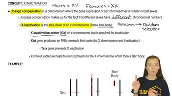
 09:30
09:30
Genotype Representation for Sex-linked Traits
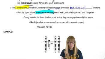 07:56
07:56Carrier Females and Phenotypic Expression
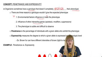 02:09
02:09From a piece of blank paper, cut out three sets of four cigar-shaped structures (a total of 12 structures). These will represent chromatids. Be sure each member of a set of four chromatids has the same length and girth. In set one, label two chromatids 'A' and two chromatids 'a.' Cut each of these chromatids about halfway across near their midpoint and slide the two 'A' chromatids together at the cuts to form a single set of attached sister chromatids. Do the same for the 'a' chromatids. In the second set of four chromatids, label two 'B' and two 'b.' Cut and slide these together as you did for the first set, joining the 'B' chromatids together and the 'b' chromatids together. Repeat this process for the third set of chromatids, labeling them as 'D' and 'd.' You now have models for three pairs of homologous chromosomes, for a total of six chromosomes. Repeat steps (h) through (l) for the alternative alignment of chromosomes you identified in step (g).
Red–green color blindness is a relatively common condition found in about 8% of males in the general population. From this, population, biologists estimate that 8% is the frequency of X chromosomes carrying a mutation of the gene encoding red and green color vision. Based on this frequency, determine the approximate frequency with which you would expect females to have red–green color blindness. Explain your reasoning.
Form a small discussion group and decide on the most likely genetic explanation for each of the following situations;
Cross A, performed by Morgan and shown in the figure below, is between a mutant male fruit fly with white eyes and a female fruit fly from a pure-breeding, red-eye stock. The figure shows that 1237 F1 progeny were produced, all of them with red eyes. In reality, this isn't entirely true. Among the 1237 F1 progeny were 3 male flies with white eyes. Give two possible explanations for the appearance of these white-eyed males.
From a piece of blank paper, cut out three sets of four cigar-shaped structures (a total of 12 structures). These will represent chromatids. Be sure each member of a set of four chromatids has the same length and girth. In set one, label two chromatids 'A' and two chromatids 'a.' Cut each of these chromatids about halfway across near their midpoint and slide the two 'A' chromatids together at the cuts to form a single set of attached sister chromatids. Do the same for the 'a' chromatids. In the second set of four chromatids, label two 'B' and two 'b.' Cut and slide these together as you did for the first set, joining the 'B' chromatids together and the 'b' chromatids together. Repeat this process for the third set of chromatids, labeling them as 'D' and 'd.' You now have models for three pairs of homologous chromosomes, for a total of six chromosomes. What are the genotypes of the daughter cells?
From a piece of blank paper, cut out three sets of four cigar-shaped structures (a total of 12 structures). These will represent chromatids. Be sure each member of a set of four chromatids has the same length and girth. In set one, label two chromatids 'A' and two chromatids 'a.' Cut each of these chromatids about halfway across near their midpoint and slide the two 'A' chromatids together at the cuts to form a single set of attached sister chromatids. Do the same for the 'a' chromatids. In the second set of four chromatids, label two 'B' and two 'b.' Cut and slide these together as you did for the first set, joining the 'B' chromatids together and the 'b' chromatids together. Repeat this process for the third set of chromatids, labeling them as 'D' and 'd.' You now have models for three pairs of homologous chromosomes, for a total of six chromosomes. Combining your work in steps (f) through (m), provide a written explanation of the connection between meiotic cell division and Mendel's law of independent assortment.