 뒤로
뒤로Chapter 4
스터디 가이드 - 스마트 노트
 자료에 맞춘 맞춤형 노트, 핵심 정의, 예시, 맥락을 확장해 제공합니다.
Overview of Eukaryotic Cells
Definition and Characteristics
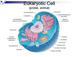
Eukaryotic cells are defined by the presence of a nucleus and membrane-bound organelles, distinguishing them from prokaryotic cells. They are found in plants, animals, fungi, and protists, and are typically larger and more complex than prokaryotes.
Nucleus: Contains genetic material (DNA) organized into multiple linear chromosomes.
Membrane-bound organelles: Include mitochondria, endoplasmic reticulum, Golgi apparatus, lysosomes, and others.
Complexity: Eukaryotic cells exhibit compartmentalization, allowing specialized functions within organelles.
The Endosymbiotic Theory
Origin of Eukaryotic Cells
The endosymbiotic theory explains the evolutionary origin of eukaryotic cells through a series of cell-merging events between ancestral eukaryotes and prokaryotes.
Mitochondria: Evolved from engulfed nonphotosynthetic prokaryotes (possibly Rickettsia species).
Chloroplasts: Evolved from engulfed photosynthetic prokaryotes (e.g., cyanobacteria).
Evidence: Mitochondria and chloroplasts have their own circular DNA, 70S ribosomes, double membranes, and replicate by binary fission.
Eukaryotic Cell Structures and Functions
Comparison with Prokaryotic Cells
Eukaryotic cells differ from prokaryotic cells in size, complexity, and the presence of organelles. They possess multiple chromosomes and undergo mitosis and meiosis for cell division.
Prokaryotes: Lack a nucleus and membrane-bound organelles; typically have a single circular chromosome.
Eukaryotes: Have a nucleus, multiple linear chromosomes, and various organelles.
Membrane-Bound Organelles
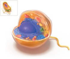
Eukaryotic cells contain specialized organelles that perform distinct functions, such as energy production, protein synthesis, and waste processing.
Mitochondria: Site of ATP production.
Endoplasmic Reticulum (ER): Rough ER synthesizes proteins; smooth ER produces lipids and detoxifies substances.
Golgi Apparatus: Modifies, sorts, and packages proteins and lipids.
Lysosomes: Contain hydrolytic enzymes for digestion.
Peroxisomes: Break down fats and amino acids, protect against toxic oxygen intermediates.
Cell Division in Eukaryotes
Mitosis and Meiosis
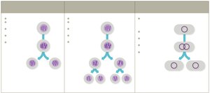
Eukaryotic cells divide by mitosis (asexual reproduction) and meiosis (sexual reproduction).
Mitosis: Produces two genetically identical diploid cells; maintains chromosome number.
Meiosis: Produces four genetically unique haploid gametes; involves two division stages and allows genetic recombination.
Binary Fission: Used by prokaryotes and organelles like mitochondria and chloroplasts; produces two identical cells.
Eukaryotic Cell Transport
Endocytosis and Exocytosis
Eukaryotic cells import and export materials via endocytosis and exocytosis, processes that require energy.
Endocytosis: Imports substances into the cell; includes pinocytosis (liquid uptake), phagocytosis (solid uptake), and receptor-mediated endocytosis.
Exocytosis: Exports substances out of the cell; vesicles fuse with the plasma membrane to release contents.
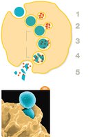
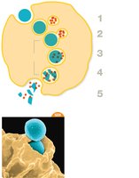
Phagocytosis
Phagocytosis is a form of endocytosis where cells engulf large particles or pathogens, important for immune defense.
Phagosome: Vesicle containing the engulfed target.
Phagolysosome: Fusion of phagosome with lysosome; hydrolytic enzymes destroy contents.
Waste expulsion: Undigested material is expelled from the cell.
Classification of Eukaryotes
Kingdoms of Eukaryotes
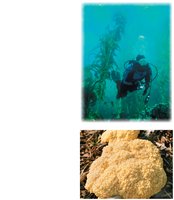
Eukaryotic organisms are classified into four kingdoms: Animals, Plants, Fungi, and Protists.
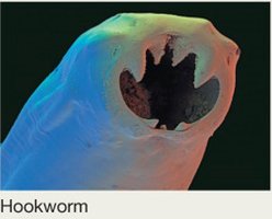
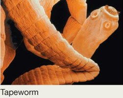
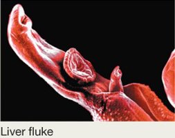
Animals: Multicellular, heterotrophic, include parasitic worms (helminths) and arthropods.
Plants: Multicellular, autotrophic, carry out photosynthesis.
Fungi: Mostly multicellular, absorb nutrients, include yeasts and molds.
Protists: Diverse group, unicellular or multicellular, autotrophic or heterotrophic.
Fungi
Structure and Classification
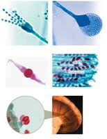
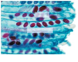
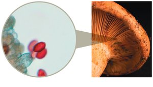
Fungi are classified based on their reproductive structures and spore types.
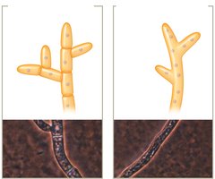
Hyphae: Tubular structures; can be septate (with divisions) or aseptate (without divisions).
Dimorphic fungi: Alternate between hyphal and yeast forms.
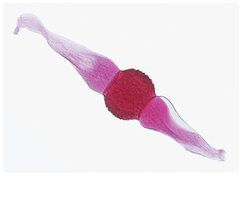
Spores: Can be asexual (conidiospores, sporangiospores) or sexual (zygospores, ascospores, basidiospores).
Fungal Diseases
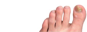
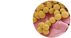
Fungi can cause diseases in humans, ranging from superficial infections to systemic illnesses.
Dermatophytes: Infect skin, hair, and nails; cause tinea infections.
Opportunistic pathogens: Infect immunocompromised hosts.
Toxins: Some fungi produce toxins (e.g., ergot toxin from Claviceps purpurea).
Protists and Protozoans
Classification and Examples
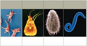
Protists are a diverse group of eukaryotes, including protozoans, algae, and slime molds. Protozoans are classified by their mode of motility.
Amoeboid: Move by pseudopodia (e.g., Entamoeba histolytica).
Flagellated: Move by flagella (e.g., Giardia lamblia).
Ciliated: Move by cilia (e.g., Tetrahymena thermophila).
Spore-forming: Move by gliding; complex life cycles (e.g., Plasmodium species).
Extracellular Structures
Plasma Membrane and Cell Wall
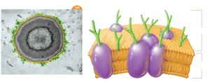
All eukaryotes have a plasma membrane, which acts as a selective barrier. Some eukaryotes have a cell wall for structural support.
Plasma membrane: Phospholipid bilayer with sterols.
Cell wall: Found in plants, fungi, and some protists; composition varies by kingdom.
Glycocalyx: Sticky extracellular layer for protection, adhesion, and communication.
Flagella and Cilia
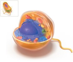
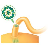
Eukaryotic flagella and cilia are used for motility and are structurally distinct from prokaryotic flagella.
Flagella: Made of tubulin, 9+2 microtubule arrangement, wave-like motion.
Cilia: Shorter and more numerous, oar-like motion.
Intracellular Structures
Ribosomes
Eukaryotic ribosomes are essential for protein synthesis and can be free in the cytoplasm or bound to the ER.
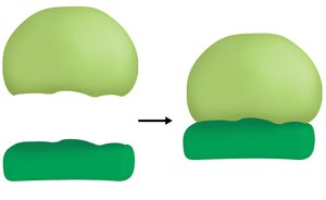
80S ribosomes: Composed of 40S and 60S subunits.
70S ribosomes: Found in mitochondria and chloroplasts; resemble prokaryotic ribosomes.
Cytoskeleton
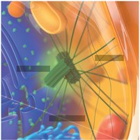
The cytoskeleton provides structural support, facilitates movement, and organizes cell contents.
Microtubules: Hollow tubes made of tubulin; form mitotic spindle.
Intermediate filaments: Rope-like fibers for tensile strength.
Microfilaments: Fine fibers made of actin; involved in contraction and movement.
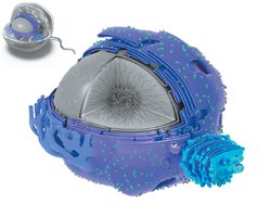
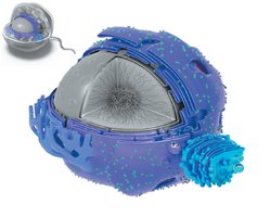
Nucleus
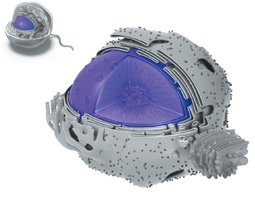
The nucleus houses DNA and is the site of transcription and ribosome assembly.
Nuclear envelope: Double membrane with nuclear pores.
Nucleolus: Site of ribosomal subunit assembly.
Chromatin: Loosely organized DNA.
Endoplasmic Reticulum (ER)
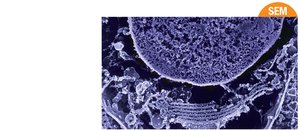
The ER is involved in protein and lipid synthesis, and detoxification.
Rough ER: Studded with ribosomes; modifies proteins.
Smooth ER: Lacks ribosomes; synthesizes lipids and detoxifies.
Golgi Apparatus
The Golgi apparatus modifies, sorts, and packages proteins and lipids for transport.
Cisternae: Disc-like, flattened sacs.
Vesicles: Transport materials between organelles and to the cell surface.
Vesicles and Vacuoles
Vesicles and vacuoles are membrane-bound sacs for storage, transport, and digestion.
Transport vesicles: Move substances within the cell.
Secretory vesicles: Discharge materials outside the cell.
Lysosomes: Digest engulfed substances.
Peroxisomes: Break down fats and amino acids.
Vacuoles: Large sacs for storage; common in plants and fungi.
Mitochondria and Chloroplasts
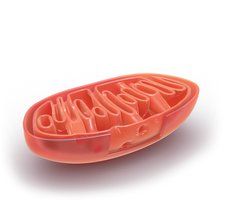
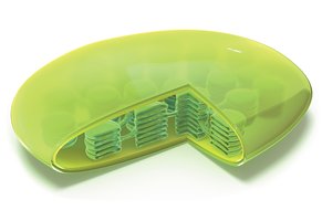
Mitochondria and chloroplasts are double-membrane organelles involved in energy production.
Mitochondria: Produce ATP, regulate cell division, and apoptosis; contain cristae and matrix.
Chloroplasts: Harvest energy from sunlight; contain thylakoids, grana, and stroma.
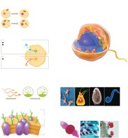
Visual Summary
Clinical Application: Case Study
Yeast and Protozoan Infections
A clinical case highlights the importance of understanding eukaryotic cell structure and function in diagnosing and treating infections. Yeast infections (e.g., Candida albicans) and protozoan infections (e.g., Trichomonas vaginalis) require different treatments due to their cellular differences.
Antibiotics: Target bacterial cells, not eukaryotic cells; thus, do not prevent yeast infections.
Antifungal drugs (e.g., fluconazole): Target ergosterol in fungal membranes, which is absent in human cells.
Protozoan drugs (e.g., metronidazole): Target specific protozoan pathways.
Key differences: Fungi and protozoans share eukaryotic features (nucleus, organelles), but differ in motility, cell wall composition, and reproductive strategies.
Microscopy: Practitioners can differentiate bacteria from yeast by size, shape, and presence of nuclei in wet mount samples.
