

Ch. 15 - Innate Immunity
Bauman - Microbiology with Diseases by Taxonomy 6th Edition
Bauman6th EditionMicrobiology with Diseases by TaxonomyISBN: 9780134832302당신이 사용하는 게 아니라요?교과서 변경
15장, 문제 2

Identify the type of hypersensitivity reaction in each photo.
<IMAGE>

검증된 단계별 안내
1
Understand that hypersensitivity reactions are classified into four main types: Type I (immediate, IgE-mediated), Type II (antibody-mediated cytotoxic), Type III (immune complex-mediated), and Type IV (delayed-type, T-cell mediated).
Examine the clinical features or histological clues in each photo carefully, noting signs such as the timing of reaction, presence of antibodies, immune complexes, or T-cell involvement.
Match the observed features in each photo to the characteristic mechanisms and manifestations of each hypersensitivity type. For example, wheal and flare reactions suggest Type I, while tissue damage with immune complexes suggests Type III.
Use knowledge of common examples: Type I includes allergies and anaphylaxis; Type II includes hemolytic anemia; Type III includes serum sickness; Type IV includes contact dermatitis.
Assign the hypersensitivity type to each photo based on the above analysis, ensuring to justify your choice by linking visual or clinical evidence to the underlying immunological mechanism.

비슷한 문제에 대한 검증된 영상 답변:

이 영상 해법은 위 문제에 도움이 된다고 튜터들이 추천한 것입니다.
영상 길이:
2m
도움이 되었나요?

주요 개념

질문에 올바르게 답하기 위해 반드시 이해해야 하는 핵심 개념들은 다음과 같습니다.

Types of Hypersensitivity Reactions

Hypersensitivity reactions are immune responses that cause tissue damage and are classified into four types: Type I (immediate, IgE-mediated), Type II (antibody-mediated cytotoxic), Type III (immune complex-mediated), and Type IV (delayed, T-cell mediated). Recognizing these types is essential for identifying the reaction based on clinical and histological features.
추천 영상:
05:48
Types of Chemical Reactions

Immunopathology and Clinical Presentation

Each hypersensitivity type has distinct pathological and clinical manifestations, such as urticaria in Type I or granuloma formation in Type IV. Understanding these presentations helps correlate the visual clues in photos with the underlying immune mechanism.
추천 영상:
04:11
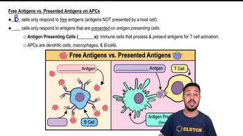
Free Antigens vs. Presented Antigens on APCs

Diagnostic Techniques in Hypersensitivity

Techniques like skin prick tests, immunofluorescence, and histological staining reveal specific immune components involved in hypersensitivity. Familiarity with these methods aids in interpreting images and confirming the type of hypersensitivity reaction shown.
추천 영상:
02:05
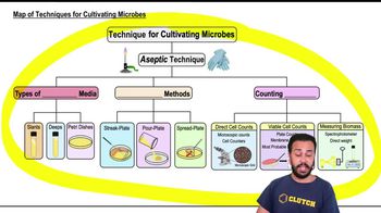
Map of Techniques for Cultivating Microbes