 뒤로
뒤로Assessment of Respiratory Function: Mini-Textbook Study Notes
스터디 가이드 - 스마트 노트
 자료에 맞춘 맞춤형 노트, 핵심 정의, 예시, 맥락을 확장해 제공합니다.
Chapter 17: Assessment of Respiratory Function |
Overview of the Respiratory System
The respiratory system is vital for oxygen transport and carbon dioxide removal, supporting cellular metabolism and life-sustaining processes. It includes structures for air conduction, gas exchange, and protection against environmental hazards.
Oxygen Transport: Oxygen is delivered to tissues for cellular respiration, while carbon dioxide is removed as a metabolic waste product.
Respiration: Involves gas exchange between atmospheric air, blood, and body cells.
Ventilation: The mechanical movement of air into and out of the lungs.
Gas Exchange: Occurs at the alveolar-capillary membrane, driven by diffusion.
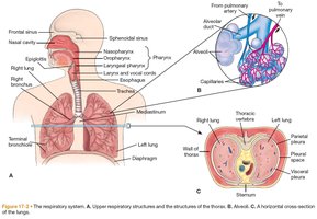
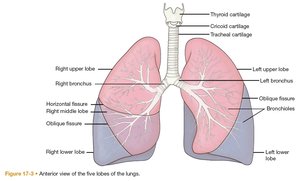
Mechanics of Ventilation
Ventilation depends on the coordinated action of the thoracic cage and diaphragm, air pressure differences, airway resistance, and lung compliance.
Inspiration: Active process requiring energy; thoracic cavity enlarges, decreasing internal pressure and drawing air in.
Expiration: Usually passive; diaphragm relaxes, increasing internal pressure and expelling air.
Airway Resistance: Influenced by airway diameter, lung volumes, and airflow velocity. Increased resistance requires more effort for ventilation.
Lung Compliance: Reflects the elasticity and expandability of the lungs and thoracic structures. Decreased compliance indicates stiffness (e.g., fibrosis); increased compliance indicates loss of recoil (e.g., emphysema).
Lung Volumes and Capacities
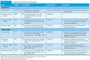
Lung function is assessed by measuring various volumes and capacities, which provide insight into respiratory health and disease states.
Volume/Capacity | Symbol | Description | Normal Value* | Significance |
|---|---|---|---|---|
Tidal Volume | VT or TV | Volume of air inhaled/exhaled with each breath | 500 mL | May vary; decreased in restrictive diseases |
Inspiratory Reserve Volume | IRV | Max air inhaled after normal inhalation | 3,000 mL | Decreased in restrictive diseases |
Expiratory Reserve Volume | ERV | Max air exhaled after normal exhalation | 1,100 mL | Decreased in obstructive diseases |
Residual Volume | RV | Air remaining after max exhalation | 1,200 mL | Increased in obstructive diseases |
Vital Capacity | VC | Max air exhaled after max inhalation (TV + IRV + ERV) | 4,600 mL | Decreased in restrictive diseases |
Inspiratory Capacity | IC | Max air inhaled after normal exhalation (TV + IRV) | 3,500 mL | Decreased in restrictive diseases |
Functional Residual Capacity | FRC | Air remaining after normal exhalation (ERV + RV) | 2,300 mL | Increased in COPD |
Total Lung Capacity | TLC | Max air in lungs after max inspiration (TV + IRV + ERV + RV) | 5,800 mL | Decreased in restrictive, increased in obstructive diseases |
Pulmonary Diffusion and Perfusion
Pulmonary diffusion is the exchange of gases at the alveolar-capillary membrane, while perfusion refers to blood flow through the pulmonary vasculature. Both are essential for effective gas exchange.
Diffusion: Facilitated by a thin membrane and large surface area.
Perfusion: Dependent on pulmonary artery pressure, gravity, and alveolar pressure.
Shunted Blood: Blood that does not participate in gas exchange due to ventilation-perfusion mismatch.
Ventilation-Perfusion (V/Q) Ratios
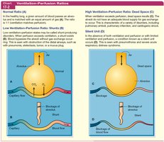
The balance between ventilation (airflow) and perfusion (blood flow) is critical for optimal gas exchange. Imbalances can lead to hypoxemia and respiratory dysfunction.
V/Q Ratio | Description | Clinical Example |
|---|---|---|
Normal | Ventilation matches perfusion | Healthy lung |
Low (Shunt) | Perfusion exceeds ventilation | Pneumonia, atelectasis |
High (Dead Space) | Ventilation exceeds perfusion | Pulmonary embolism |
Silent Unit | Both ventilation and perfusion are decreased | ARDS, pneumothorax |
Comprehensive Assessment of Respiratory Function
Health History and Common Symptoms
A thorough health history and symptom assessment are essential for identifying respiratory disease and guiding further evaluation.
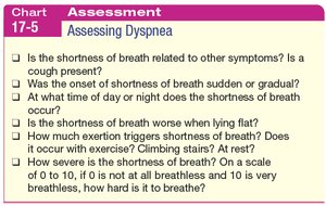
Dyspnea: Subjective sensation of breathing discomfort; may be acute or chronic, and associated with various conditions (e.g., COPD, heart failure, asthma).
Cough: Reflex to clear airways; characteristics (timing, quality, associated sputum) provide diagnostic clues.
Sputum Production: Nature and color may indicate infection, chronic disease, or malignancy.
Chest Pain: May be pulmonary, cardiac, musculoskeletal, or anxiety-related; requires careful assessment of onset, quality, and relation to breathing.
Wheezing: High-pitched, musical sound indicating airway narrowing.
Hemoptysis: Coughing up blood; may result from infection, malignancy, or vascular abnormalities.
Physical Examination
Physical assessment includes inspection, palpation, percussion, and auscultation of the respiratory system.
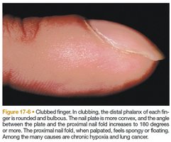
General Appearance: Observe for clubbing of fingers (chronic hypoxia), cyanosis, and use of accessory muscles.
Clubbing: Rounded, bulbous distal phalanges; associated with chronic hypoxia and lung disease.
Assessment Techniques
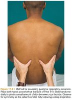
Respiratory Excursion: Assesses range and symmetry of thoracic expansion by observing thumb movement during deep inspiration and expiration.
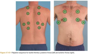
Tactile Fremitus: Palpation of chest wall vibrations as patient speaks; altered in various lung conditions.
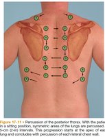
Percussion: Tapping on the chest wall to assess underlying tissue density (resonant, dull, or hyperresonant sounds).
Age-Related Changes in the Respiratory System
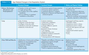
Aging affects the structure and function of the respiratory system, increasing susceptibility to disease and reducing reserve capacity.
Component | Structural Changes | Functional Changes | Clinical Findings |
|---|---|---|---|
Defense Mechanisms | Decreased cilia, mucus, and cough reflex | Impaired clearance, increased infection risk | Cough, infection, abnormal breath sounds |
Lung | Loss of alveolar surface area, decreased elasticity | Reduced gas exchange, increased V/Q mismatch | Decreased PaO2, increased residual volume |
Chest Wall/Muscles | Calcification, decreased muscle strength | Reduced chest expansion, increased work of breathing | Kyphosis, barrel chest, shallow breathing |
Diagnostic Evaluation of Respiratory Function
Pulmonary Function Tests (PFTs)
PFTs are used to assess lung volumes, ventilatory function, and gas exchange, aiding in diagnosis, monitoring, and preoperative evaluation. Results are interpreted based on deviation from predicted normal values, considering patient demographics.
Arterial and Venous Blood Gas Studies
Arterial Blood Gas (ABG): Measures oxygenation, ventilation, and acid-base status; obtained via arterial puncture.
Venous Blood Gas (VBG): Provides information on tissue oxygen extraction; less invasive than ABG.
Pulse Oximetry
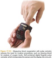
Pulse oximetry is a noninvasive method for monitoring oxygen saturation (SpO2) of hemoglobin. It is widely used in clinical and home settings for rapid assessment of oxygenation.
Normal SpO2: >95%
Values <90%: Indicate inadequate oxygenation
Limitations: Does not detect hyperoxemia, affected by poor perfusion, nail polish, and skin pigmentation
Other Diagnostic Methods
End-Tidal CO2 (ETCO2) Monitoring: Noninvasive measurement of exhaled CO2; useful for monitoring ventilation and confirming airway placement.
Cultures and Sputum Studies: Identify pathogens and malignant cells; guide therapy for respiratory infections.
Imaging Studies: Chest x-ray, CT, MRI, and nuclear scans provide structural and functional information about the lungs and thorax.
Endoscopic Procedures: Bronchoscopy, thoracoscopy, and thoracentesis allow direct visualization, sampling, and intervention in the respiratory tract.
Summary Table: Key Physical Findings in Common Respiratory Diseases
Physical findings such as breath sounds, percussion notes, and tactile fremitus help differentiate between normal and pathological states (e.g., pneumonia, COPD, pleural effusion).
Additional info: This guide integrates foundational respiratory physiology and clinical assessment principles relevant to introductory psychology and allied health students, emphasizing the interplay between structure, function, and diagnostic reasoning.
