 Back
BackAxial Skeleton: Structure and Components
Study Guide - Smart Notes
 Tailored notes based on your materials, expanded with key definitions, examples, and context.
Axial Skeleton
Overview of the Axial Skeleton
The axial skeleton forms the central axis of the human body, providing support and protection for the brain, spinal cord, and thoracic organs. It consists of the skull, vertebral column, and thoracic cage.
Skull: Protects the brain and forms the structure of the face.
Vertebral column: Supports the head and body, protects the spinal cord.
Thoracic cage: Protects the heart and lungs, supports the shoulder girdles and upper limbs.
Classification of Bones by Shape
Types of Bones
Bones are classified according to their shapes, which relate to their functions:
Long bones: Longer than they are wide; have a shaft and two ends (e.g., humerus).
Short bones: Cube-shaped; found in the wrist and ankle.
Sesamoid bones: Develop within tendons (e.g., patella).
Flat bones: Thin, flattened, and often curved (e.g., sternum, most skull bones).
Irregular bones: Complicated shapes (e.g., vertebrae, hip bones).
Sutural bones: Small bones found within the sutures of the skull.
Skull
Structure and Components
The skull consists of the cranial bones, which protect the brain, and the facial bones, which form the structure of the face, including the upper and lower jaws, nose, and orbits.
Cranial bones (8): Occipital, frontal, sphenoid, ethmoid, two parietal, two temporal.
Facial bones (14): Maxilla (2), lacrimal (2), nasal (2), zygomatic (2), mandible, palatine (2), inferior nasal conchae (2), vomer.
Sutures are immovable joints that connect the cranial bones, including the sagittal, lambdoid, squamous, and coronal sutures.
Anterior View of the Skull
The anterior view of the skull displays the bones forming the forehead, orbits (eye sockets), nasal cavity, nasal septum, and upper and lower jaws.
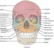
Key Features of the Skull
Hard palate: Formed by the maxillary and palatine bones.
TMJ (temporomandibular joint): The only movable joint of the face, between the mandible and temporal bone.
Teeth: Anchored in the mandible (lower jaw) and maxillary bones (upper jaw).
Special Bones and Structures
Sphenoid bone: Forms part of the cranial floor; contains the sella turcica, which houses the pituitary gland.
Ethmoid bone: Located at the midline; forms part of the nasal septum and the medial wall of the orbit.
Nasal septum: Formed by the perpendicular plate of the ethmoid bone and the vomer; septal cartilage completes the structure.
Conchae: Curved bones (superior, middle, and inferior) that form air passages in the nasal cavity, aiding in air flow and filtration.
Sinuses
Paranasal Sinuses
Sinuses are air-filled cavities within certain skull bones. They lighten the skull, add resonance to the voice, and drain into the nasal cavity.
Frontal sinus: Pain in the forehead if infected.
Ethmoid sinus: Pain between the eyes if infected.
Maxillary sinus: Pain in the upper jaw if infected.
Sphenoid sinus: Pain in the back of the eye if infected.
Hyoid Bone
Unique Features
The hyoid bone does not articulate with any other bone. It is held in place by ligaments and provides attachment for muscles of the mouth, tongue, and larynx. The epiglottis and pharynx are located posterior to it.
Vertebral Column
Structure and Regions
The vertebral column consists of 24 vertebrae, the sacrum, and the coccyx. It is divided into three main regions:
Cervical (C1–C7): Neck region.
Thoracic (T1–T12): Upper back, articulates with ribs.
Lumbar (L1–L5): Lower back, supports most body weight.
The sacrum is formed by the fusion of five sacral vertebrae, and the coccyx by the fusion of four small coccygeal vertebrae.
Curvatures of the Spine
Primary curvatures: Thoracic and sacrococcygeal (present at birth).
Secondary curvatures: Cervical and lumbar (develop after birth).
Abnormal Curvatures
Scoliosis: Abnormal lateral curvature.
Kyphosis: Excessive thoracic curvature.
Lordosis: Excessive lumbar curvature.
Structure of a Typical Vertebra
Body: Weight-bearing portion.
Vertebral arch: Formed by pedicles and laminae.
Processes: Transverse, spinous, superior and inferior articular processes.
Vertebral foramen: Passage for the spinal cord.
Intervertebral foramen: Passage for spinal nerves.
Intervertebral discs: Fibrocartilaginous pads (anulus fibrosus and nucleus pulposus) between vertebrae, allowing movement and absorbing shock.
Special Vertebrae
Atlas (C1): Supports the skull; allows nodding "yes" motion.
Axis (C2): Has the dens; allows rotation "no" motion.
Thoracic Vertebrae and Rib Articulation
Thoracic vertebrae have facets for rib attachment.
Ribs articulate with both the vertebral body and transverse process.
Herniated Disc
Weakening of the anulus fibrosus can cause the nucleus pulposus to protrude, compressing spinal nerves and causing pain or muscle weakness.
Thoracic Cage
Structure and Function
The thoracic cage protects the heart and lungs and supports the upper limbs. It consists of the sternum, ribs, and thoracic vertebrae.
Sternum: Flat bone with three parts: manubrium, body, xiphoid process.
Ribs: 12 pairs; all attach posteriorly to the thoracic vertebrae.
True ribs (1–7): Attach directly to the sternum via costal cartilages.
False ribs (8–12): Do not attach directly to the sternum; ribs 8–10 merge with cartilage, ribs 11–12 are floating ribs with no anterior attachment.
Articulations of the Axial Skeleton
Key Joints and Movements
Atlas (C1) and occipital condyle: Allows nodding "yes" motion.
Atlas (C1) and axis (C2): Allows rotation "no" motion.
L5 and sacrum: Articulation between lumbar spine and sacrum.
Sacrum and coccyx: Articulation at the base of the spine.
Sacrum and ilium: Forms part of the pelvic girdle.
Newborn Skull
Fontanelles and Growth
The bones of the newborn skull are separated by fontanelles, which are areas of fibrous connective tissue. These allow for growth of the skull after birth. The facial bones are small and underdeveloped at birth, and the mastoid process is not yet formed.
