 Back
BackCardiology and the Cardiovascular System: Medical Terminology Study Guide
Study Guide - Smart Notes
 Tailored notes based on your materials, expanded with key definitions, examples, and context.
Cardiology and the Cardiovascular System
Introduction to Cardiology
Cardiology is the medical specialty focused on the anatomy, physiology, diseases, and treatments of the cardiovascular system. Understanding medical terminology in this field is essential for accurate communication and patient care.
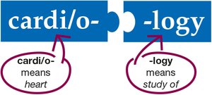
Cardi/o-: means heart
-logy: means study of
Cardiologist: a physician specializing in heart and blood vessel disorders
Structures of the Cardiovascular System
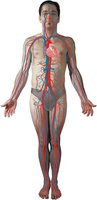
The cardiovascular system consists of the heart and blood vessels, forming a continuous circuit that delivers blood throughout the body.
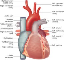
Heart: muscular organ located in the thoracic cavity, behind the sternum and between the lungs
Blood Vessels: arteries, veins, and capillaries that transport blood
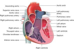
Anatomy of the Heart
External and Internal Features
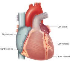
The heart is roughly the size of a fist and contracts at least once per second. Its external surface is irregular, with mounds and grooves reflecting internal chambers and containing fat, blood vessels, and nerves.
Membranes and Layers
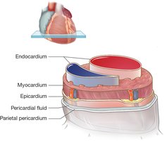
The heart is surrounded by a two-layered membrane called the pericardium. The outer layer is the parietal pericardium, and the inner layer is the epicardium. Pericardial fluid fills the space between these layers, reducing friction during heartbeats. The myocardium is the muscular layer responsible for contraction, and the endocardium lines the chambers and valves.
Heart Chambers
The heart has four chambers:
Atria: two small upper chambers (right and left)
Ventricles: two large lower chambers (right and left)
Septum: central wall dividing the heart into right and left sides
Heart Valves
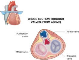
Four valves control blood flow:
Tricuspid valve: between right atrium and right ventricle
Pulmonary valve: between right ventricle and pulmonary arteries
Mitral valve: between left atrium and left ventricle
Aortic valve: between left ventricle and aorta
The chordae tendineae are ropelike strands that help keep the tricuspid and mitral valves closed during ventricular contraction. Heart sounds "lubb-dupp" are produced by the opening and closing of these valves.
Thoracic Cavity and Mediastinum
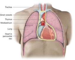
The heart is located in the mediastinum, a central area within the thoracic cavity. The mediastinum also contains the great vessels, esophagus, trachea, and thymus.
Blood Vessels
Types and Functions
Blood vessels are vascular channels lined with endothelium. There are three main types:
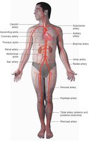
Arteries: carry blood away from the heart
Capillaries: connect arteries and veins, deliver blood to cells
Veins: carry blood toward the heart
Arteries
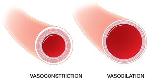
Arteries branch into arterioles and carry oxygen-rich blood. They have smooth muscle for vasoconstriction and vasodilation, which regulate blood pressure.
Capillaries
Capillaries are the smallest blood vessels, allowing single-file passage of blood cells and exchange of gases, nutrients, and waste.
Veins
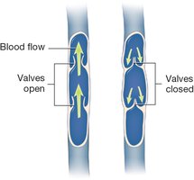
Veins and venules carry oxygen-poor blood to the heart. Many veins have valves to ensure one-way flow and prevent backflow due to gravity.
Circulation Pathways
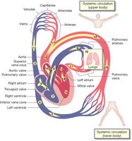
Blood circulates through two main pathways:
Systemic circulation: vessels throughout the body except the lungs
Pulmonary circulation: vessels to, within, and from the lungs
Blood passes through both pathways in one complete trip:
Deoxygenated blood from the body enters the right atrium via the superior and inferior vena cava
Blood moves to the right ventricle, then through the pulmonary valve to the pulmonary arteries and lungs
In the lungs, blood releases CO2 and picks up O2
Oxygenated blood returns via pulmonary veins to the left atrium
Blood moves to the left ventricle, then through the aortic valve to the aorta and systemic circulation
Physiology of the Heart
Conduction System
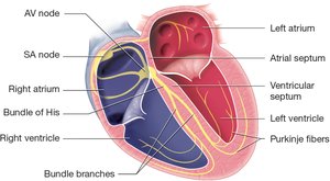
The heart's conduction system initiates and transmits electrical impulses:
Sinoatrial (SA) node: pacemaker, initiates heartbeat
Atrioventricular (AV) node: receives impulse from SA node
Bundle of His and bundle branches: transmit impulse through septum
Purkinje fibers: cause ventricles to contract
Heart Rhythm and Rate
Systole: contraction phase
Diastole: relaxation phase
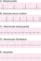
Normal sinus rhythm (NSR): SA node controls heart rate
Ectopic sites: abnormal rhythm if other areas produce impulses
Nervous System Control
Parasympathetic division: releases acetylcholine, maintains resting heart rate
Sympathetic division: releases norepinephrine and epinephrine, increases heart rate during activity and stress
Common Cardiovascular Diseases
Heart Diseases
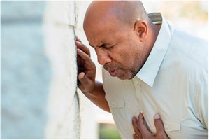
Angina pectoris: chest pain due to reduced blood flow to the heart
Congestive heart failure: heart's inability to pump effectively, causing edema
Myocardial infarction: heart attack, death of heart tissue due to lack of oxygen
Blood Vessel Diseases
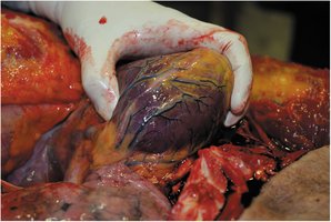
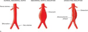
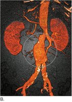
Aneurysm: abnormal bulging of an artery wall
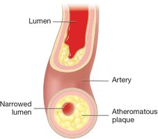
Atherosclerosis: buildup of plaque in arteries, narrowing the lumen
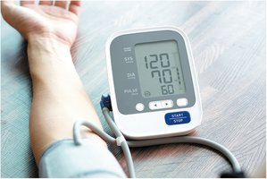
Hypertension: high blood pressure
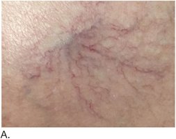
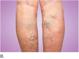
Varicose veins: enlarged, twisted veins, often in the legs
Conduction System Diseases
Arrhythmias: abnormal heart rhythms, including bradycardia, tachycardia, fibrillation, and asystole
Laboratory and Diagnostic Procedures
Exercise Stress Tests
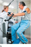
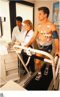
Bicycle and treadmill stress tests: monitor heart function during exercise
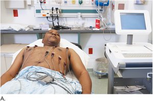
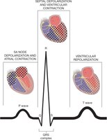
Electrocardiography (ECG/EKG)
Precordial chest leads: electrodes placed on chest
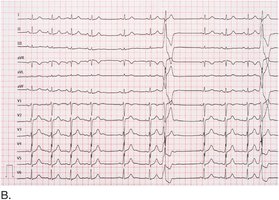
12-lead ECG: comprehensive heart electrical activity recording
ECG tracing: shows P wave, QRS complex, and T wave
Ultrasonography and Echocardiography
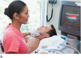
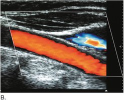
Color flow duplex ultrasonography: visualizes blood flow and velocity
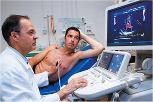
Echocardiography: uses sound waves to create images of heart structures
Medical Procedures
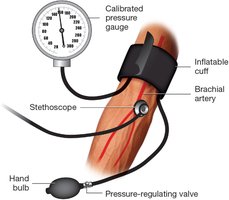
Blood Pressure Measurement
Sphygmomanometer: device for measuring blood pressure
Systolic pressure: force of ventricular contraction
Diastolic pressure: resting pressure in the artery
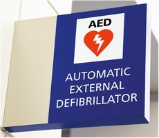
Automatic External Defibrillator (AED)
AED: device used to restore normal heart rhythm in emergencies
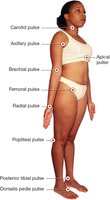
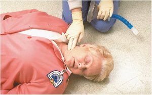
Pulse Points
Pulse points: locations where arterial pulse can be felt
Used to assess heart rate and blood flow
Drugs and Surgical Procedures
Cardiovascular Drugs
Digitalis drugs: derived from the foxglove plant (Digitalis purpurea), used to treat heart conditions

Surgical Procedures
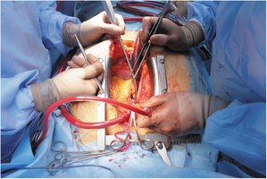
Open heart surgery: sternum is split to access the heart
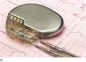
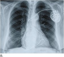
Pacemaker implantation: device placed under skin to regulate heart rhythm
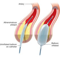
Balloon angioplasty: catheter with balloon compresses plaque in arteries
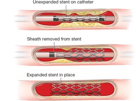
Stent placement: expanded stent keeps artery open
Valve replacement: artificial valve implanted to restore function
Summary Table: Heart Chambers and Valves
Chamber | Valve | Location |
|---|---|---|
Right Atrium | Tricuspid Valve | Between right atrium and right ventricle |
Right Ventricle | Pulmonary Valve | Between right ventricle and pulmonary arteries |
Left Atrium | Mitral Valve | Between left atrium and left ventricle |
Left Ventricle | Aortic Valve | Between left ventricle and aorta |
Key Equations
Blood Pressure Formula:
Cardiac Output Formula:
Additional info:
Scientific names and terminology are expanded for clarity.
Tables and equations are provided for academic completeness.
