 Back
Study Guide: Hematology and Immunology – Blood and Lymphatic System
Study Guide - Smart Notes
 Tailored notes based on your materials, expanded with key definitions, examples, and context.
Hematology and Immunology
Overview
Hematology is the medical specialty focused on the anatomy and physiology of blood, while immunology deals with the lymphatic system and the body's immune response. These systems are essential for transporting nutrients, defending against pathogens, and maintaining homeostasis.
Anatomy of the Blood
Hematopoiesis
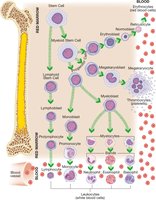
Hematopoiesis is the process of blood cell formation, occurring in the red marrow of long and flat bones. All blood cells originate from stem cells and mature through various stages before entering the bloodstream.
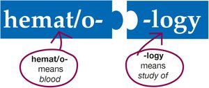
hemat/o-: means blood
-poiesis: means formation
Stem cells differentiate into myeloid and lymphoid lineages, producing erythrocytes, leukocytes, and thrombocytes.
Erythrocytes (Red Blood Cells)
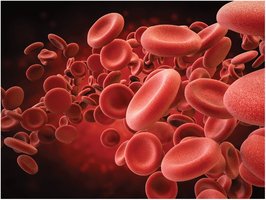
Erythrocytes are the most numerous blood cells, responsible for transporting oxygen and carbon dioxide via hemoglobin. They lack a nucleus, cannot divide, and have a lifespan of about 120 days.
Produced as erythroblasts, released as reticulocytes
Removed by the spleen, which recycles iron and amino acids
Production stimulated by erythropoietin when levels are low
Leukocytes (White Blood Cells)
Leukocytes are divided into granulocytes and agranulocytes, each with distinct functions in immunity.
Granulocytes: Neutrophils, Eosinophils, Basophils
Agranulocytes: Lymphocytes, Monocytes
Neutrophils
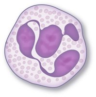
Most common leukocyte (54-62%), granulocyte with neutral-staining granules and multi-lobed nucleus. They are phagocytes, ingesting bacteria.
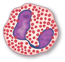
Eosinophils
Granulocytes (1-3%), granules stain pink with eosin, nucleus has two lobes. They release chemicals to destroy foreign cells and parasites.
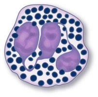
Basophils
Least common granulocyte (0.5-1%), granules stain blue with hematoxylin, multi-lobed nucleus. They release histamine to increase blood flow.
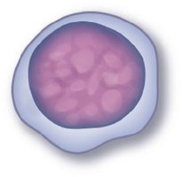
Lymphocytes
Agranulocytes (25-33%), round nucleus, little cytoplasm. Includes B cells (produce antibodies) and T cells (cell-mediated immunity).
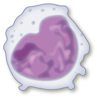
Monocytes
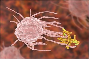
Largest leukocyte (3-7%), kidney-shaped nucleus, few granules. They mature into macrophages and perform phagocytosis.
Thrombocytes (Platelets)
Thrombocytes are cell fragments essential for blood clotting. They originate from megakaryocytes in the bone marrow.
thromb/o-: blood clot
-cyte: cell
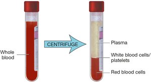
Plasma
Plasma is the liquid component of blood, making up 55% of its volume. It transports nutrients, wastes, and suspends formed elements.
Contains albumin, electrolytes, and clotting factors
Serum is plasma without clotting factors
Anatomy of the Lymphatic System
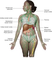
Lymphatic Vessels and Nodes
Lymphatic vessels begin as capillaries, collect tissue fluid (lymph), and transport it to lymph nodes for filtration. Lymph nodes are grouped in areas prone to infection or cancer.
Lymphoid Tissues and Organs
Lymphoid tissues (tonsils, adenoids, Peyer patches, appendix) and organs (thymus, spleen) are involved in immune responses. The thymus matures T lymphocytes, and the spleen recycles erythrocytes and stores blood.
Physiology of Blood Clotting
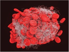
Clotting Process
Blood clotting involves vessel constriction, platelet aggregation, and activation of clotting factors. Fibrin strands form a mesh that traps erythrocytes, creating a clot.
Clotting factors: fibrinogen, prothrombin (liver), thromboplastin (tissue), calcium, others
Heparin limits clot size
Immune Response
Allergic Reaction
An allergy is a hypersensitivity to allergens, causing local or systemic reactions. Severe systemic reactions can lead to anaphylaxis.

Leukocyte Functions
Leukocytes coordinate the immune response by attacking pathogens and cancer cells. Neutrophils and monocytes are phagocytes, while eosinophils and basophils release chemicals to destroy invaders.
Lymphocyte Types
NK (Natural Killer) cells: destroy cancer and viral-infected cells
B cells: produce antibodies
T cells: cytotoxic (kill infected cells), helper (stimulate response), memory (remember pathogens), suppressor (limit response)
Immune Response Physiology
Monocytes engulf pathogens and release chemicals
Interferon stimulates antiviral substances and NK cells
Interleukin stimulates B, T, and NK cells, produces fever
Tumor necrosis factor destroys endotoxins and cancer cells
Antibodies coat pathogens, attract leukocytes, and mark for destruction
Complements drill holes in pathogen cell walls
Blood Diseases
Erythrocyte Diseases
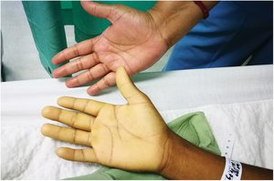
Anemia: Reduced erythrocyte count or hemoglobin, causing pallor and fatigue
Iron deficiency anemia: Microcytic, hypochromic erythrocytes
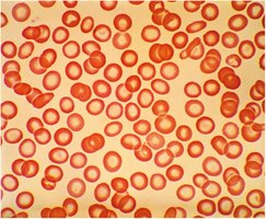
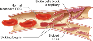
Sickle cell anemia: Abnormal, sickled erythrocytes block capillaries, cause pain and clots
Leukocyte Diseases
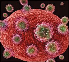
HIV: Attacks helper T cells, impairs immune function
Leukemia: Overproduction of immature leukocytes
Thrombocyte and Blood Clotting Disorders
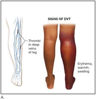
Deep Vein Thrombosis (DVT): Blood clot in deep vein, causes erythema, warmth, swelling
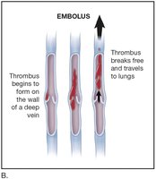
Embolus: Thrombus breaks free, travels to lungs
Lymphatic System Disorders
Lymphadenopathy: Enlarged lymph nodes due to infection or cancer
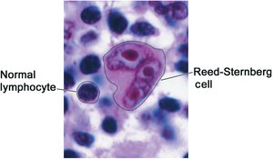
Hodgkin lymphoma: Presence of Reed-Sternberg cells

Laboratory and Medical Procedures
Blood Cell Tests
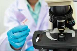
Blood smears are used to examine blood cells under a microscope for diagnostic purposes.
Blood Chemistry Analysis
Automated analyzers perform multiple blood tests to assess chemical and cellular components.

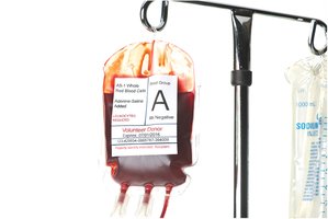
Blood Transfusion
Blood is collected from donors, stored, and transfused to patients as needed. One unit equals 500 mL (about 1 pint).
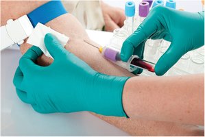
Phlebotomy
Phlebotomy is the process of drawing blood for laboratory testing, typically using a tourniquet and vacuum tube.
Summary Table: Blood Cell Types
Cell Type | Appearance | Function |
|---|---|---|
Erythrocyte | Red, biconcave, no nucleus | Transport O2 and CO2 |
Neutrophil | Granules, multi-lobed nucleus | Phagocytosis of bacteria |
Eosinophil | Pink granules, two-lobed nucleus | Destroy parasites, allergic response |
Basophil | Blue granules, multi-lobed nucleus | Release histamine |
Lymphocyte | Round nucleus, little cytoplasm | Antibody production, cell-mediated immunity |
Monocyte | Large, kidney-shaped nucleus | Phagocytosis, become macrophages |
Thrombocyte | Cell fragment | Blood clotting |
Key Medical Terminology
hemat/o-: blood
erythr/o-: red
leuk/o-: white
thromb/o-: clot
-cyte: cell
-poiesis: formation
Important Equations
Blood volume calculation:
Hematocrit calculation:
Additional info: Academic context was added to clarify cell functions, disease mechanisms, and laboratory procedures for completeness.
