Skip to main content
Microbiology
My Course
Learn
Exam Prep
AI Tutor
Study Guides
Textbook Solutions
Flashcards
Explore
Try the app
My Course
Learn
Exam Prep
AI Tutor
Study Guides
Textbook Solutions
Flashcards
Explore
Try the app
Back
Introduction to Microscopes quiz #1
You can tap to flip the card.
Which type of microscope has the best resolution?
You can tap to flip the card.
👆
Which type of microscope has the best resolution?
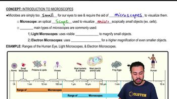
Electron microscopes have the best resolution, allowing visualization of extremely small objects like viruses and molecules.
Track progress
Control buttons has been changed to "navigation" mode.
1/40
Related flashcards
Related practice
Recommended videos
Introduction to Microscopes quiz #2
Introduction to Microscopes
40 Terms
Introduction to Microscopes quiz #3
Introduction to Microscopes
9 Terms
Introduction to Microscopes definitions
Introduction to Microscopes
15 Terms
Introduction to Microscopes
9. Microscopes
5 problems
Topic
Monica
Magnification, Resolution, & Contrast
9. Microscopes
7 problems
Topic
Nicole
9. Microscopes - Part 1 of 2
6 topics
12 problems
Chapter
Nicole
9. Microscopes - Part 2 of 2
8 topics
10 problems
Chapter
Nicole
Guided course
06:13
Introduction to Microscopes
3392
views
65
rank
1
comments
Terms in this set (40)
Hide definitions
Which type of microscope has the best resolution?
Electron microscopes have the best resolution, allowing visualization of extremely small objects like viruses and molecules.
Who developed the first single-lens microscope?
Anton van Leeuwenhoek developed the first single-lens microscope.
Why does the sample on a microscope slide need to be very thin?
A sample needs to be thin so that light can pass through it, allowing clear visualization under a light microscope.
What are the magnification abilities of each of the objective lenses on a compound light microscope?
Objective lenses typically provide magnifications such as 4x (scanning), 10x (low power), 40x (high power), and sometimes 100x (oil immersion).
Which microscope uses a series of lenses to magnify an object in steps?
A compound light microscope uses a series of lenses to magnify objects in steps.
Through which microscope were cells first observed?
Cells were first observed through a light microscope.
Where are the focus controls on a microscope located?
Focus controls are typically located on the side of the microscope, near the stage.
What is the maximum magnification of most classroom compound light microscopes?
Most classroom compound light microscopes have a maximum magnification of about 400x to 1000x.
What is the advantage of viewing a specimen at 40x as opposed to at 400x magnification?
At 40x, you can see a larger area of the specimen, making it easier to locate features before zooming in for detail at higher magnification.
Which type of microscope is usually used to examine viruses?
Electron microscopes are usually used to examine viruses.
Which microscope achieves the highest magnification and greatest resolution?
Electron microscopes achieve the highest magnification and greatest resolution.
Angela would like to look at living algae from pond water. What type of equipment should she use?
Angela should use a light microscope to observe living algae.
You have a sample and put it under your light microscope. What can you see?
You can see cells, bacteria, and other objects large enough for light microscope resolution, but not viruses.
You look at crossed threads under the microscope. This allows you to find out what?
Looking at crossed threads helps determine the depth of field and which thread is on top or bottom.
What microscope would be appropriate to generate a 3-dimensional image of a bacterium?
A scanning electron microscope (SEM) is appropriate for generating 3-dimensional images of bacteria.
Which type of microscope is best for observing the presence or absence of trichomes?
A light microscope is best for observing the presence or absence of trichomes.
Indicate the correct order of steps as you bring an object into focus under high power.
Start with low power objective, focus using coarse adjustment, switch to high power, and use fine adjustment to sharpen the image.
A device for spinning a specimen at high speed until it separates into its component parts is a what?
A centrifuge is used to spin specimens at high speed for separation.
Which part of an optical microscope is the platform on which the specimen slide rests?
The stage is the platform where the specimen slide rests.
When using the high power objective, only the fine adjustment knob should be used.
True. Only the fine adjustment knob should be used with the high power objective.
What serves as a handle for carrying the microscope?
The arm of the microscope serves as a handle for carrying.
What type of microscope most likely produced the image of a mitochondrion?
An electron microscope most likely produced the image of a mitochondrion.
The visualization of bacteria requires the use of a compound microscope.
True. A compound light microscope is commonly used to visualize bacteria.
A specimen that is spread thinly across a slide in order to be viewed under a microscope is called a what?
A smear is a specimen spread thinly across a slide.
What holds a microscope slide in position?
Stage clips hold the microscope slide in position.
When preparing a wet mount specimen for viewing, the specimen should be covered with what?
The specimen should be covered with a cover slip.
The optical microscopes used in physicians' office laboratories usually have __________ objectives.
They usually have three or four objectives: scanning, low, high, and sometimes oil immersion.
The scanning, low, and high power objectives are mounted on the:
They are mounted on the revolving nosepiece.
What controls the amount of light allowed on the specimen?
The diaphragm controls the amount of light on the specimen.
What items do you need to make a wet mount?
You need a slide, cover slip, specimen, and water or another liquid.
Which microscope achieves the highest magnification and greatest resolution?
Electron microscopes achieve the highest magnification and greatest resolution.
When focusing a specimen, you should always start with the _____________ objective.
You should always start with the lowest power (scanning) objective.
Select the components used by a bright-field microscope to view a specimen.
Bright-field microscopes use a light source, condenser, objective lenses, and ocular lens.
What holds a specimen while being viewed under the microscope?
The stage and stage clips hold the specimen.
The iris of the optical microscope is __________.
The iris is a diaphragm that regulates the amount of light passing through the specimen.
What is the part of the microscope that connects the head to the base?
The arm connects the head to the base.
When focusing a specimen you should always start with what objective?
You should start with the scanning (lowest power) objective.
Which part of the bright-field microscope focuses light on the specimen?
The condenser focuses light on the specimen.
Which setting would allow you to observe a specimen with the highest possible magnification?
Using the highest power objective lens (e.g., 100x oil immersion) and the ocular lens.
You should always carry the microscope by the
You should carry the microscope by the arm and support the base.
 BackBack
 06:13
06:13