Skip to main content
Microbiology
My Course
Learn
Exam Prep
AI Tutor
Study Guides
Textbook Solutions
Flashcards
Explore
Try the app
My Course
Learn
Exam Prep
AI Tutor
Study Guides
Textbook Solutions
Flashcards
Explore
Try the app
Back
Introduction to Microscopes quiz #2
You can tap to flip the card.
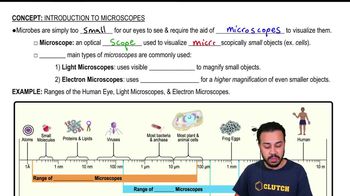
The compound light microscope is most useful for viewing
You can tap to flip the card.
👆
The compound light microscope is most useful for viewing
It is most useful for viewing cells, bacteria, and larger microorganisms.
Track progress
Control buttons has been changed to "navigation" mode.
1/40
Related flashcards
Related practice
Recommended videos
Introduction to Microscopes quiz #1
Introduction to Microscopes
40 Terms
Introduction to Microscopes quiz #3
Introduction to Microscopes
9 Terms
Introduction to Microscopes definitions
Introduction to Microscopes
15 Terms
Introduction to Microscopes
9. Microscopes
5 problems
Topic
Monica
Magnification, Resolution, & Contrast
9. Microscopes
7 problems
Topic
Nicole
9. Microscopes - Part 1 of 2
6 topics
12 problems
Chapter
Nicole
9. Microscopes - Part 2 of 2
8 topics
10 problems
Chapter
Nicole
Guided course
06:13
Introduction to Microscopes
3392
views
65
rank
1
comments
Terms in this set (40)
Hide definitions
The compound light microscope is most useful for viewing
It is most useful for viewing cells, bacteria, and larger microorganisms.
What is the lens that is attached to the nosepiece?
The objective lens is attached to the nosepiece.
When presenting, which tool would you use to magnify a section of the slide?
You would use the microscope's objective lenses to magnify sections of the slide.
What type of microscope produces colored images that appear three-dimensional?
A confocal microscope produces colored, three-dimensional images.
The microscope field is the ________.
The microscope field is the area visible through the eyepiece.
If you use a compound light microscope, a 2-μm bacterial cell is best seen at which magnification?
A 2-μm bacterial cell is best seen at 1000x magnification.
You begin your observations using the ________ lens and the ________ adjustment knob.
You begin with the scanning (lowest power) lens and the coarse adjustment knob.
What is the objective lens used to locate the specimen?
The scanning objective lens (usually 4x) is used to locate the specimen.
Blank is the area you see through the microscope.
Field of view is the area you see through the microscope.
The advantage of a compound-light microscope over an electron microscope is that it
It allows observation of living specimens and is less expensive and easier to use.
An electron microscope would be the best choice for viewing
An electron microscope is best for viewing viruses, proteins, and very small structures.
The microscope best for viewing living cells at low levels of magnification is the
The light microscope is best for viewing living cells at low magnification.
Adjust the stage to center the sample in the field of view.
Use the stage controls to move the sample and center it in the field of view.
Which microscope produces a three-dimensional image of the object under the lens?
A scanning electron microscope (SEM) produces a three-dimensional image.
One advantage of electron microscopes over light microscopes is their
Electron microscopes have much higher magnification and resolution.
Which structure is best observed using a compound light microscope?
Cells and most bacteria are best observed using a compound light microscope.
Organelles, granules, and bacterial endospores are best observed using which microscope?
A compound light microscope is best for observing organelles, granules, and bacterial endospores.
The low power objective magnifies a specimen by:
The low power objective typically magnifies by 10x.
What holds the specimen when you use a microscope?
The stage and stage clips hold the specimen.
Samples for a compound light microscope are typically prepared on _______.
Samples are prepared on glass slides.
The visualization of viruses requires the use of a(n) microscope.
Visualization of viruses requires the use of an electron microscope.
One primary advantage of light microscopy over electron microscopy is that
Light microscopy allows observation of living specimens.
Which lenses in a microscope are magnifying lenses?
The objective lenses and the ocular lens are magnifying lenses.
Observing microorganisms through a microscope
Allows visualization of cells and microbes too small for the naked eye.
Why do you place one hand under the base of the microscope as you carry it?
To support the weight and prevent dropping the microscope.
What is the lens at the top of the microscope through which the specimen is viewed?
The ocular lens (eyepiece) is at the top of the microscope.
Always carry a microscope using both hands.
True. Always use both hands to carry a microscope safely.
When using the microscope, how is the focus adjusted?
Focus is adjusted using the coarse and fine adjustment knobs.
True or false: Darkfield microscopes show light images on a dark background.
True. Darkfield microscopes show light images on a dark background.
What term is used to describe the image produced by the ocular lens of a microscope?
The term is virtual image.
The _____ microscope uses multiple glass lenses to help distinguish details of thickness.
The compound light microscope uses multiple glass lenses.
Which type of microscope achieves the greatest resolution and highest magnification?
Electron microscopes achieve the greatest resolution and highest magnification.
You should begin viewing a specimen with what objective lens?
You should begin with the scanning (lowest power) objective lens.
The ______ microscope is used to visualize most types of bacteria.
The compound light microscope is used to visualize most types of bacteria.
When storing the microscope, the stage should be left in the ____ position.
The stage should be left in the lowest position.
The objective lenses of the compound light microscope are attached to the
They are attached to the revolving nosepiece.
What does it mean to have parfocal objectives?
Parfocal objectives stay in focus when switching between lenses with minimal adjustment.
What was the significance of the invention of the microscope in 1666?
It allowed scientists to visualize and study microscopic organisms and cells for the first time.
Which type of microscope does not use light in forming the specimen image?
Electron microscopes do not use light; they use electrons.
What is the function of the revolving nosepiece on a microscope?
The revolving nosepiece holds objective lenses and allows switching between them.
 BackBack
 06:13
06:13