 Back
BackIntroduction to Microbiology Study Guide
 03:36
03:36

Terms in this set (29)
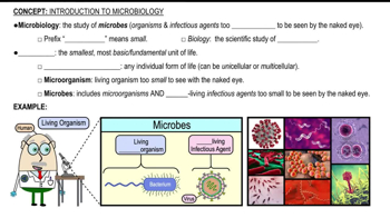
Microbiology is the study of microbes, which are microscopic organisms including bacteria, viruses, fungi, and protozoa.
A microbe is a microscopic organism that can be prokaryotic or eukaryotic, cellular or noncellular.
Prokaryotic organisms lack a nucleus and membrane-bound organelles; eukaryotic organisms have a nucleus and organelles.
Cellular microbes include bacteria, archaea, fungi, and protozoa; noncellular microbes include viruses and prions.
Pathogens cause disease in healthy hosts; opportunistic pathogens cause disease only when the host's defenses are compromised.
Redi's experiment showed that maggots on decaying meat came from fly eggs, disproving spontaneous generation.
To demonstrate that microbes come from the environment, supporting biogenesis and disproving spontaneous generation.
The germ theory states that microorganisms are the cause of many diseases.
1. Microbe found in diseased but not healthy hosts.
2. Microbe isolated and grown in pure culture.
3. Microbe causes disease when introduced to healthy host.
4. Microbe reisolated from experimentally infected host.
Aseptic technique prevents contamination and reduces healthcare-associated infections by sterilizing instruments and using hand hygiene.
Observation, question, hypothesis, experiment, analysis, and conclusion.
Observation is noting facts; interpretation is explaining or drawing conclusions from observations.
Taxonomy classifies and names organisms to organize biological diversity.
Bacteria, Archaea, and Eukarya.
Genus name capitalized, species lowercase, both italicized or underlined (e.g., Escherichia coli).
A strain is a genetic variant or subtype of a microbial species, e.g., E. coli K-12 or O157:H7.
Mutualism, commensalism, and parasitism.
Normal microbiota are microbes that normally live on or in the human body without causing disease.
Disruption can lead to overgrowth of harmful microbes and disease; caused by antibiotics, illness, or environmental changes.
A biofilm is a community of microbes attached to a surface, embedded in extracellular polymeric substances (EPS).
Pure culture contains one microbial species; mixed culture contains multiple species.
Staining increases contrast to better visualize microbes under a microscope.
Basic dyes are positively charged and stain cells; acidic dyes are negatively charged and stain the background.
To differentiate bacteria into Gram-positive (purple) and Gram-negative (pink) based on cell wall structure.
To identify bacteria with waxy cell walls rich in mycolic acid, such as Mycobacterium species.
Bright-field uses light passing through the specimen; dark-field illuminates specimen against a dark background for better contrast.
Immersion oil reduces light refraction, increasing resolution and clarity at high magnifications.
TEM passes electrons through specimens for internal structures (2D images); SEM scans surface for 3D images.
A technique using fluorescent-labeled antibodies to detect specific microbes under a fluorescence microscope.