 Back
BackMicrobiology Microscopy and Taxonomy Basics
 06:13
06:13Terms in this set (19)
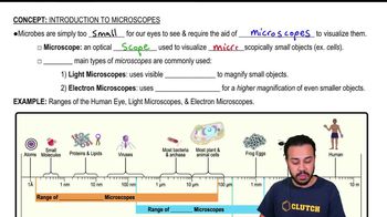
Microscopy is the use of a microscope to view objects that cannot be seen with the naked eye.
Electromagnetic radiation provides the light or electron beams used in microscopy to illuminate specimens for visualization.
Resolving power depends on the wavelength of the radiation used and the numerical aperture of the microscope lens.
Contrast is enhanced by staining, which colors specimens to distinguish structures more clearly under the microscope.
Bright-field uses light passing through the specimen; dark-field uses scattered light to view specimens against a dark background; phase microscopy enhances contrast by amplifying differences in refractive index.
TEM transmits electrons through thin specimens to show internal structures; SEM scans surface electrons to produce 3D surface images. TEM offers higher resolution; SEM shows surface details.
A smear spreads microorganisms on a slide to create a thin layer for microscopic examination.
Heat fixation kills microorganisms, adheres them to the slide, and preserves their shape for staining.
Chemical fixation preserves cellular structures by using chemicals to stabilize and fix specimens on slides.
Acidic dyes stain the background or negatively charged components, useful for negative staining techniques.
Basic dyes stain the positively charged components of cells, such as nucleic acids and proteins, highlighting microorganisms.
Simple stains use one dye to color all cells similarly; differential stains use multiple dyes to distinguish cell types or structures.
Taxonomy organizes and classifies organisms to understand relationships and provide universal names.
Microorganisms reproduce asexually and exchange genes horizontally, making species boundaries unclear and classification challenging.
The hierarchy is: Domain, Kingdom, Phylum, Class, Order, Family, Genus, Species.
Binomial nomenclature assigns each organism a two-part Latin name: genus (capitalized) and species (lowercase), e.g., Escherichia coli.
Spread bacteria thinly on a slide, air dry, then pass the slide briefly through a flame to fix cells.
Cover a heat-fixed smear with a basic dye, let sit briefly, rinse with water, and blot dry before viewing.
Gram-positive bacteria retain crystal violet stain appearing purple; gram-negative bacteria do not and appear pink/red after counterstaining.