 Back
BackEmbryogenesis and the Human Body Plan: Foundations for Nutrition and Development
Study Guide - Smart Notes
 Tailored notes based on your materials, expanded with key definitions, examples, and context.
Basic Body Plan of Adult Humans
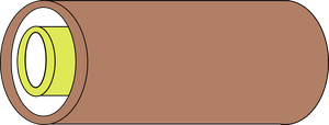
Tube-in-a-Tube Structure
The human body is organized as a tube within a tube, a fundamental anatomical arrangement that supports digestion, circulation, and other vital functions. - Outer Tube: Represents the body wall, including skin, muscles, and bones. - Inner Tube: Represents the digestive tract (gut tube), which runs from mouth to anus. - Coelom: The space between the tubes, allowing organs to expand and move (e.g., heart beating, lung expansion, food passage). 
Serous Membranes
Serous membranes line and protect internal body cavities and organs. - Parietal Serosa: Lines the inside of the body wall. - Visceral Serosa: Covers the outside of organs. - Peritoneum: The serous membrane associated with the abdominal cavity, crucial for organ protection and movement. 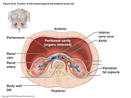
Vertebrate Structures
The human body plan shares features with other vertebrates, including a spinal cord, notochord, and segmented muscles. - Spinal Cord: Central nervous system structure running along the dorsal body wall. - Kidneys: Retroperitoneal organs located laterally to the midline. 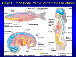
Fertilization
Sperm Transport and Egg Meeting
Fertilization is the process where sperm and egg unite, initiating human development. - Location: Occurs in the ampulla of the fallopian tube, a dilated region optimal for fertilization. - Sperm Journey: Sperm travel approximately 180 mm (~7 inches), which is about 3600 times their own length, to reach the egg. - Union: Only one sperm successfully penetrates the egg, triggering embryogenesis. 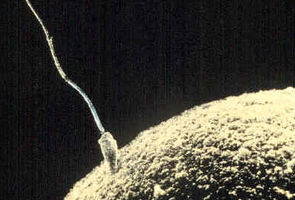
Early Embryonic Development (Embryogenesis)
Cleavage and Formation of Multicellular Embryo
After fertilization, the zygote undergoes rapid cell divisions called cleavage. - Cleavage: Series of mitotic divisions without cell growth, increasing cell number and surface-to-volume ratio. - Zygote: The single cell formed by fertilization. - Embryo: The developing organism from 2-8 weeks post-fertilization.
Morula and Blastocyst Formation
- Morula: A solid ball of cells, typically formed when the embryo has more than 16 cells. - Blastocyst: A hollow ball of cells with a fluid-filled cavity. - Trophoblast: The outer layer of the blastocyst, which contributes to placenta formation. - Inner Cell Mass: The group of cells inside the blastocyst that will develop into the embryo proper.
Implantation
The blastocyst implants into the uterine wall, a critical step for pregnancy. - Endometrium: The mucous membrane lining the uterus, which nourishes the blastocyst before implantation. - Trophoblast Function: Binds to the endometrium, causing an inflammatory response and facilitating implantation. - Implantation Process: The blastocyst burrows deeper into the endometrium, which grows over it to secure the embryo.
Summary Table: Key Stages of Early Human Development
Stage | Description | Key Features |
|---|---|---|
Zygote | Single cell formed by fertilization | Genetic material from both parents |
Cleavage | Rapid cell division without growth | Increased cell number, same size |
Morula | Solid ball of cells (>16 cells) | No cavity, compact structure |
Blastocyst | Hollow ball with fluid-filled cavity | Trophoblast (outer), inner cell mass |
Implantation | Blastocyst embeds in uterine wall | Endometrium, trophoblast interaction |
Additional info:
- Understanding embryogenesis is essential for nutrition students, as early development is highly sensitive to maternal nutrition, micronutrient status, and metabolic health. - The tube-in-a-tube body plan is foundational for digestive system structure and function, which is central to nutritional science.
