 Back
BackOptical Instruments: Cameras, Human Eye, Vision Correction, Magnifiers, and Microscopes
Study Guide - Smart Notes
 Tailored notes based on your materials, expanded with key definitions, examples, and context.
Optical Instruments
Cameras
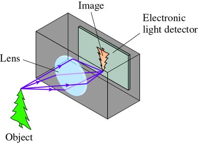
Cameras are devices that project a real image onto a plane surface, where it can be recorded onto film or an electronic detector. The basic principle involves a converging lens focusing light from an object onto a detector, such as a charge-coupled device (CCD). The sharpness and exposure of the image are controlled by the lens position, aperture size, and shutter speed.
Pinhole Camera: Uses a small hole to allow only one light ray from each point of an object, limiting light and image quality.
Standard Camera: Uses a converging lens to project a real image onto an electronic detector.
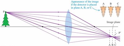
Focusing: Achieved by moving the lens so the detector is at the image plane.
Aperture: The iris diaphragm controls the diameter d of the lens opening, affecting exposure and depth of field.
f-number: Defined as the ratio of lens focal length f to lens diameter d:
Shutter Speed: Controls exposure time, typically from 1/1000 s to 1/30 s.
Example: The image below shows how a camera lens projects light rays from an object onto an electronic detector, forming a real image.
The next image illustrates how the position of the detector relative to the image plane affects image sharpness.
The Human Eye
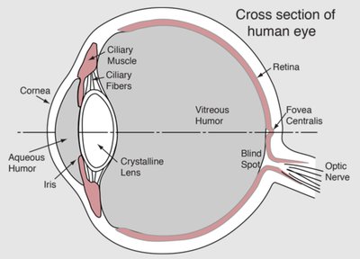
The human eye is a biological optical instrument that refracts incoming light to form an image on the retina. The cornea, aqueous humor, and lens work together to focus light, while the iris controls the amount of light entering, similar to a camera diaphragm. The retina acts as the light-sensitive detector.
Cornea: Provides most of the eye's refractive power due to its curvature and refractive index difference with air.
Lens: Adjusts refractive power via shape changes controlled by ciliary muscles (accommodation).
Iris: Regulates light entry.
Retina: Receives the focused image.
Accommodation: The process by which the eye changes lens shape to focus on near or far objects.
Near Point: Closest distance for clear vision (~25 cm for normal vision).
Far Point: Farthest distance for clear vision (essentially infinity for normal vision).
Vision Defects and Correction
Two common vision defects are myopia (nearsightedness) and hyperopia (farsightedness). These are caused by abnormal eye shape or lens system strength and are corrected using lenses.
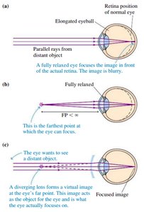
Myopia (Nearsightedness)
Cause: Elongated eyeball or overly curved cornea; image forms in front of retina.
Correction: Use a diverging lens to form a virtual image at the eye's far point, allowing the eye to focus on distant objects.
Thin Lens Equation:
Optical Power: (in diopters, D)
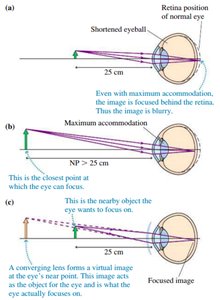
Hyperopia (Farsightedness)
Cause: Shortened eyeball or weak lens system; image forms behind retina.
Correction: Use a converging lens to form a virtual image at the eye's near point, allowing the eye to focus on close objects.
Magnifiers (Simple Magnifying Lens)
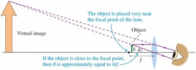
A magnifier is a converging lens used to enlarge the apparent size of an object. The object is placed just inside the focal point, producing a virtual image farther away than the near point. The angular magnification is determined by the ratio of the angles subtended by the image and the object at the eye.
Angular Magnification:
Small-Angle Approximation: , , so
Example: The image below shows the near point of the eye and the angle subtended by an object.
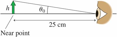
The next image illustrates how a magnifying lens creates a virtual image, increasing the angle subtended at the eye.
Microscopes
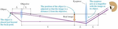
A microscope uses two lenses (objective and eyepiece) to achieve high magnification. The object is placed just outside the focal point of the objective, which forms a real, enlarged, inverted image. The eyepiece then acts as a magnifier for this image.
Objective Lens: Forms a real image at distance L (tube length) from the objective.
Eyepiece: Magnifies the real image.
Magnification of Objective:
Magnification of Eyepiece:
Overall Magnification:
Example Calculation
Given: Objective focal length cm, eyepiece focal length cm, lens separation cm.
Tube Length: cm
Object Distance: cm
Objective Magnification:
Eyepiece Magnification:
Overall Magnification:
Additional info: The notes provide a stepwise method for analyzing two-lens systems, which is essential for understanding compound optical instruments like microscopes.
