 Back
BackOptical Instruments: The Eye, Vision Correction, and the Microscope
Study Guide - Smart Notes
 Tailored notes based on your materials, expanded with key definitions, examples, and context.
Chapter 25: Optical Instruments
Introduction to Optical Instruments
Optical instruments utilize the principles of mirrors and lenses to manipulate light and form images for various applications. These devices are essential in both daily life and specialized fields such as medicine and scientific research.
Everyday Examples: Cameras, eyeglasses, binoculars
Medical Examples: Endoscopes (internal examination), ophthalmoscopes (eye examination)
This chapter focuses on the physics behind cameras, projectors, the human eye, and reviews magnifiers, microscopes, and telescopes.
The Eye as an Optical Instrument
Structure and Function of the Eye
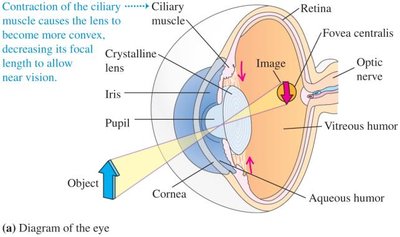
The human eye is a complex optical system that forms real images of objects on the retina through refraction at the cornea and lens. For clear vision, the image must be focused precisely on the retina.
Cornea and Lens: Responsible for most of the eye's focusing power.
Retina: Light-sensitive layer where the image is formed.
Accommodation: The eye adjusts the shape of the lens to focus on objects at different distances.
Far Point: The most distant point at which the eye can see clearly.
Near Point: The closest point at which the eye can see clearly.
Normal Vision
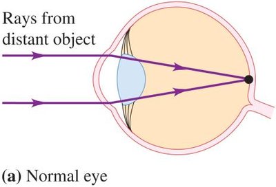
In a normal eye, parallel rays from a distant object are focused directly on the retina, resulting in a sharp image.
Vision Defects and Correction
Common vision defects include hyperopia (farsightedness) and myopia (nearsightedness). These conditions are corrected using lenses with appropriate focal lengths and powers.
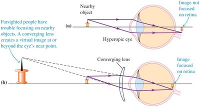
Hyperopia (Farsightedness): The image forms behind the retina. Corrected with a converging lens (positive focal length).
Myopia (Nearsightedness): The image forms in front of the retina. Corrected with a diverging lens (negative focal length).
Lens Power: Defined as the reciprocal of the focal length (in meters). The unit is the diopter (D):
where is the power in diopters and is the focal length in meters.
Correction of Hyperopia (Farsightedness)
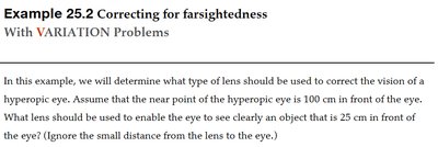
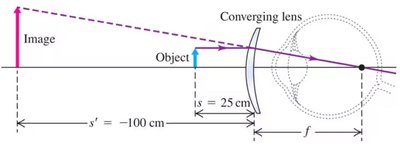
Farsighted people have difficulty focusing on nearby objects. A converging lens creates a virtual image at or beyond the eye's near point, allowing the eye to focus the image on the retina.
Example 25.2: Determining the lens needed to correct a hyperopic eye with a near point at 100 cm, to see an object at 25 cm.
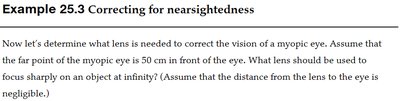
Correction of Myopia (Nearsightedness)
Nearsighted people cannot focus on distant objects. A diverging lens is used to focus parallel rays from distant objects onto the retina.
The Microscope
Principle and Construction
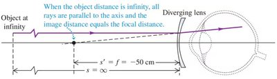
A microscope is used to achieve high magnification, allowing observation of very small objects. It consists of two main lenses: the objective and the eyepiece.
Objective Lens: Placed close to the object; forms a real, enlarged, and inverted image just beyond its focal point.
Eyepiece (Ocular): Acts as a magnifier for the image formed by the objective, producing a virtual, further enlarged image.
The total magnification () of a compound microscope is the product of the lateral magnification of the objective () and the angular magnification of the eyepiece ():
where and are the focal lengths of the objective and eyepiece, is the image distance from the objective, and is the object distance from the objective.
Example: Observing Bacteria
Example 25.5: Calculating the required magnification to observe bacteria with a compound microscope. For an eyepiece with 15x magnification, the objective must provide sufficient magnification to achieve a total of 2000x. The size of a half-micron bacterium is compared to the period at the end of a sentence.
Summary Table: Vision Defects and Correction
Condition | Image Location | Correction | Type of Lens |
|---|---|---|---|
Normal Vision | On retina | None needed | — |
Hyperopia (Farsightedness) | Behind retina | Move image forward | Converging (positive) |
Myopia (Nearsightedness) | In front of retina | Move image backward | Diverging (negative) |
Additional info: The examples and diagrams provided reinforce the application of lens formulas and ray diagrams in understanding and correcting vision defects, as well as the construction and use of microscopes in scientific observation.
