 Back
BackA Tour of the Cell: Structure, Function, and Methods of Study
Study Guide - Smart Notes
 Tailored notes based on your materials, expanded with key definitions, examples, and context.
Cells: The Fundamental Units of Life
Introduction to Cell Theory
Cells are the basic unit of structure and function in all living organisms. They can exist as single-celled organisms or as part of multicellular organisms, such as plants and animals. The cell theory states that all living things are composed of cells, and all cells arise from pre-existing cells.
Unicellular organisms (e.g., Paramecium) consist of a single cell that performs all life functions.
Multicellular organisms (e.g., plants, animals) are composed of many specialized cells.
All cells contain DNA as their genetic material.
Cell Theory Key Points
All living organisms are composed of cells.
The cell is the simplest collection of matter that can be alive.
Cells are related by their descent from earlier cells.
Cells share common features but can differ substantially in structure and function.
Studying Cells: Microscopy and Biochemistry
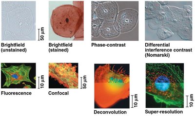
Microscopy: Visualizing Cells
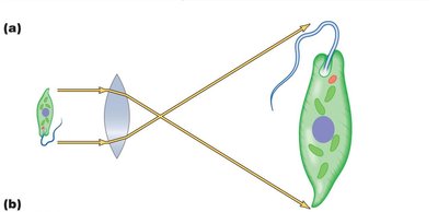
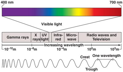
Cells are typically too small to be seen with the naked eye. Microscopes are essential tools for visualizing cells and their components. There are two main types of microscopy: light microscopy and electron microscopy.
Light Microscopy (LM): Uses visible light and glass lenses to magnify images of specimens.
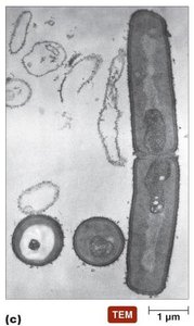
Electron Microscopy (EM): Uses electron beams and magnetic lenses for much higher resolution.
Parameters of Microscopy
Magnification: The ratio of an object's image size to its real size.
Resolution: The minimum distance two points can be separated and still be distinguished as two points.
Contrast: Differences in intensity between parts of the sample, often enhanced by staining.
Types of Light Microscopy
Brightfield (unstained and stained): Standard illumination; staining increases contrast but usually kills cells.
Phase-contrast and Differential Interference Contrast (Nomarski): Enhance contrast in unstained cells.
Fluorescence Microscopy: Uses fluorescent stains and UV light to visualize specific cell components.
Confocal and Super-resolution Microscopy: Provide sharper, three-dimensional images.
Electron Microscopy
Scanning Electron Microscope (SEM): Provides 3D images of specimen surfaces.
Transmission Electron Microscope (TEM): Reveals internal structures by passing electrons through thin sections.
Cryo-Electron Microscopy (cryo-EM): Preserves specimens at very low temperatures for imaging in near-native states.
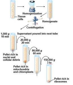
Cell Fractionation
Cell fractionation is a technique used to separate cellular components based on size and density using centrifugation. This allows scientists to study the functions of individual organelles.
Differential centrifugation: Sequentially spins cell homogenates at increasing speeds to separate nuclei, mitochondria, and ribosomes.
Cell Types: Prokaryotic vs. Eukaryotic Cells
Three Domains of Life
Bacteria (prokaryotes)
Archaea (prokaryotes)
Eukarya (eukaryotes: protists, fungi, animals, plants)
Prokaryotic Cells
Lack a nucleus; DNA is in an unbound region called the nucleoid.
No membrane-bound organelles.
Generally smaller than eukaryotic cells.
Eukaryotic Cells
DNA is contained within a nucleus bounded by a double membrane.
Possess membrane-bound organelles (e.g., mitochondria, ER, Golgi apparatus).
Generally larger and more complex than prokaryotic cells.
Comparison Table: Prokaryotic vs. Eukaryotic Cells
Feature | Prokaryotic Cells | Eukaryotic Cells |
|---|---|---|
Nucleus | Absent | Present |
Membrane-bound organelles | Absent | Present |
Size | Smaller | Larger |
DNA location | Nucleoid (unbound) | Nucleus (bound by envelope) |
Cell wall | Usually present, no cellulose | Present in plants (cellulose), absent in animals |
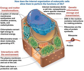
Internal Organization of Eukaryotic Cells
Compartmentalization by Internal Membranes
Eukaryotic cells have internal membranes that divide the cell into compartments, allowing specialized functions to occur efficiently. The plasma membrane controls the passage of materials in and out of the cell, and the surface area-to-volume ratio limits cell size.
Biological Membranes
Composed mainly of a double layer of phospholipids and proteins.
Phospholipids have hydrophilic heads and hydrophobic tails, forming a bilayer.
The Nucleus and Ribosomes
Nucleus
Contains most of the cell's DNA.
Enclosed by a double membrane (nuclear envelope) with nuclear pores for molecular transport.
Contains the nucleolus, the site of ribosomal RNA (rRNA) synthesis.
DNA is organized into chromosomes, composed of chromatin (DNA + proteins).
Ribosomes
Complexes of rRNA and protein; sites of protein synthesis.
Free ribosomes function in the cytosol; bound ribosomes are attached to the rough ER or nuclear envelope.
The Endomembrane System
Components and Functions
Nuclear envelope
Endoplasmic reticulum (ER): Rough ER (protein synthesis), Smooth ER (lipid synthesis, detoxification, Ca2+ storage)
Golgi apparatus: Modifies, sorts, and packages proteins and lipids
Lysosomes: Digestive compartments containing hydrolytic enzymes
Vacuoles: Storage, waste disposal, water balance, and plant cell growth
Plasma membrane
Endoplasmic Reticulum (ER)
Rough ER: Studded with ribosomes; synthesizes proteins and glycoproteins, distributes transport vesicles, and produces membranes.
Smooth ER: Lacks ribosomes; synthesizes lipids, detoxifies drugs, and stores calcium ions.
Golgi Apparatus
Consists of flattened sacs (cisternae).
Modifies ER products, manufactures macromolecules, sorts and packages materials into vesicles.
Lysosomes
Membranous sacs of hydrolytic enzymes that digest macromolecules.
Involved in phagocytosis and autophagy (recycling cell components).
Vacuoles
Large vesicles with diverse functions: food vacuoles (phagocytosis), contractile vacuoles (water balance), central vacuole (plant cell growth and storage).
Energy Conversion: Mitochondria and Chloroplasts
Mitochondria
Sites of cellular respiration; generate ATP using oxygen.
Double membrane structure; inner membrane folded into cristae for increased surface area.
Contain their own DNA and ribosomes.
Chloroplasts
Sites of photosynthesis in plants and algae.
Contain chlorophyll, thylakoids (stacked into grana), and stroma (internal fluid).
Have their own DNA and ribosomes.
Peroxisomes
Specialized metabolic compartments; produce hydrogen peroxide and convert it to water.
Involved in fatty acid breakdown and detoxification (e.g., in liver cells).
Endosymbiont Theory
Mitochondria and chloroplasts are thought to have originated from prokaryotic cells engulfed by ancestral eukaryotes, forming a symbiotic relationship. Evidence includes their double membranes, own DNA, and ribosomes.
The Cytoskeleton
Structure and Function
The cytoskeleton is a network of protein fibers that organizes cell structure and activities, providing support, shape, and facilitating movement.
Microtubules: Thickest; made of tubulin; involved in cell shape, organelle movement, and chromosome separation.
Microfilaments (Actin Filaments): Thinnest; made of actin; support cell shape, involved in muscle contraction and cell motility.
Intermediate Filaments: Middle diameter; provide mechanical support and anchor organelles.
Extracellular Components and Cell Junctions
Extracellular Matrix (ECM) in Animal Cells
Composed of glycoproteins (collagen, proteoglycans, fibronectin).
ECM proteins bind to integrins in the plasma membrane, influencing cell behavior and gene activity.
Cell Walls in Plants
Provide protection, shape, and prevent excessive water uptake.
Composed mainly of cellulose.
Contain multiple layers: primary cell wall, middle lamella, and sometimes a secondary cell wall.
Cell Junctions
Tight junctions: Prevent leakage of extracellular fluid (animal cells).
Desmosomes: Anchor cells together (animal cells).
Gap junctions: Allow communication between cells (animal cells).
Plasmodesmata: Channels between plant cells for transport and communication.
Integration of Cellular Components
No single component of the cell works alone; cellular functions are the result of coordinated activities among organelles, the cytoskeleton, and the plasma membrane. For example, the destruction of bacteria by macrophages involves the cytoskeleton, lysosomes, and plasma membrane working together.
