 Back
BackA Tour of the Cell: Structure, Function, and Organization
Study Guide - Smart Notes
 Tailored notes based on your materials, expanded with key definitions, examples, and context.
Cell Structure and Organization
Introduction to Cells
Cells are the fundamental units of life, forming the basis of all living organisms. The study of cell structure and function reveals how life is organized at the microscopic level and how cellular components contribute to the overall physiology of organisms.
Cell Theory: All living things are composed of cells; cells are the smallest units of life; all cells arise from pre-existing cells.
Cell Diversity: Cells vary in size, shape, and function, but share common structural features.
Evolutionary Origin: Cells are related by descent from earlier cells, with chemical evolution occurring 4–5 billion years ago.
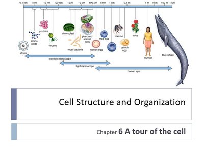
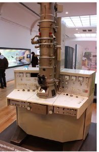
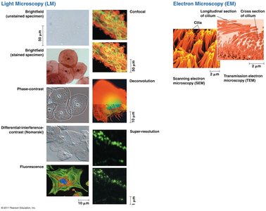
Microscopy and Cell Observation
Microscopy has enabled the discovery and study of cells, allowing scientists to observe structures invisible to the naked eye. Different types of microscopes provide varying levels of resolution and magnification.
Light Microscopy: Uses visible light to observe living or stained cells; resolution limit ~200 nm.
Electron Microscopy: Uses electron beams for much higher resolution (~2 nm); includes Transmission EM (internal structures) and Scanning EM (surface details).
Key Parameters: Magnification (enlargement), Resolution (clarity), and Contrast (distinguishing features).
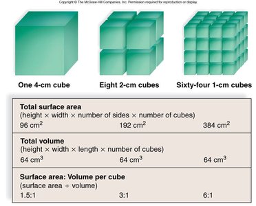
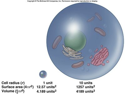
Cell Size and Surface Area-to-Volume Ratio
The size of cells is constrained by the need to efficiently exchange materials with their environment. The surface area-to-volume ratio is a critical factor in determining cell size and function.
Surface Area-to-Volume Ratio (SA:V): As a cell grows, its volume increases faster than its surface area, limiting the rate of material exchange.
Small Cells: Have a higher SA:V ratio, facilitating efficient transport of substances.
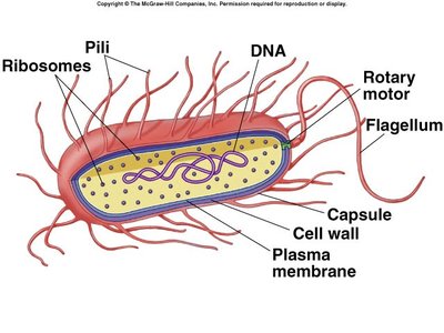
Prokaryotic and Eukaryotic Cells
Prokaryotic Cells
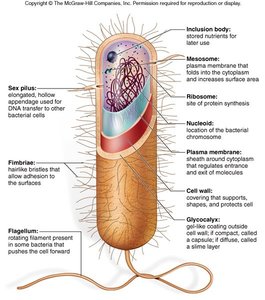
Prokaryotic cells are simpler and smaller than eukaryotic cells, lacking a true nucleus and membrane-bound organelles. They include bacteria and archaea.
Genetic Material: DNA is located in the nucleoid region, not enclosed by a membrane.
Cell Wall: Provides structural support; composed of peptidoglycan in bacteria.
Other Features: Ribosomes (protein synthesis), plasma membrane, capsule (protection), flagella (movement), pili/fimbriae (adhesion).

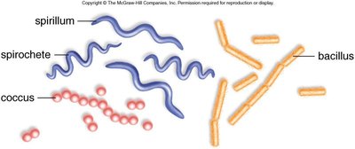
Shapes of Prokaryotes
Coccus: Spherical
Bacillus: Rod-shaped
Spirillum/Spirochete: Spiral-shaped
Eukaryotic Cells
Eukaryotic cells are more complex, containing a true nucleus and various membrane-bound organelles. They include protists, fungi, plants, and animals.
Nucleus: Contains genetic material (DNA) enclosed by a nuclear envelope.
Organelles: Specialized structures such as mitochondria, endoplasmic reticulum, Golgi apparatus, lysosomes, and (in plants) chloroplasts.
Cytoskeleton: Provides structural support and facilitates movement.
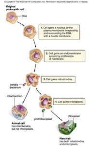
Endosymbiotic Theory
The endosymbiotic theory explains the origin of mitochondria and chloroplasts as formerly free-living prokaryotes engulfed by ancestral eukaryotic cells.
Evidence: Double membranes, circular DNA, and 70S ribosomes in mitochondria and chloroplasts.
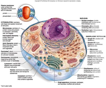
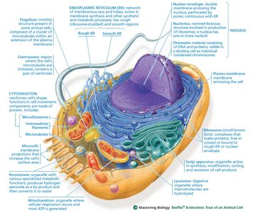
Cellular Components and Their Functions
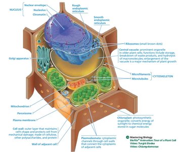
Nucleus
The nucleus is the control center of the cell, housing genetic material and coordinating activities such as growth and reproduction.
Chromatin: DNA-protein complex; condenses to form chromosomes during cell division.
Nucleolus: Site of ribosomal RNA synthesis.
Nuclear Envelope: Double membrane with pores for molecular transport.
Ribosomes
Ribosomes are the sites of protein synthesis, found either free in the cytoplasm or bound to the endoplasmic reticulum.
Structure: Composed of rRNA and proteins; two subunits (large and small).
Types: 70S in prokaryotes and organelles; 80S in eukaryotic cytoplasm.
Endomembrane System
The endomembrane system is a network of membranes involved in synthesis, modification, and transport of cellular materials.
Components: Nuclear envelope, endoplasmic reticulum (ER), Golgi apparatus, lysosomes, vacuoles, and plasma membrane.
Vesicles: Transport materials between organelles.
Endoplasmic Reticulum (ER)
Rough ER: Studded with ribosomes; synthesizes proteins for secretion or membrane insertion.
Smooth ER: Lacks ribosomes; synthesizes lipids, detoxifies chemicals, stores calcium ions.
Golgi Apparatus
The Golgi apparatus modifies, sorts, and packages proteins and lipids for delivery to various destinations.
Structure: Flattened membranous sacs called cisternae.
Lysosomes and Vacuoles
Lysosomes: Contain hydrolytic enzymes for digestion of macromolecules and recycling of cellular components (autophagy).
Vacuoles: Storage organelles; central vacuole in plants stores water, ions, and waste products.
Mitochondria and Chloroplasts
Mitochondria: Sites of cellular respiration; generate ATP from organic fuels using oxygen.
Chloroplasts: Sites of photosynthesis in plants and algae; contain chlorophyll and other pigments.
Peroxisomes
Peroxisomes are metabolic compartments that break down fatty acids and detoxify harmful substances, producing hydrogen peroxide as a byproduct, which is then converted to water.
Cytoskeleton
The cytoskeleton is a dynamic network of protein filaments that provides structural support, facilitates cell movement, and organizes organelles.
Microtubules: Hollow tubes of tubulin; involved in cell shape, organelle movement, and cell division.
Microfilaments: Actin filaments; support cell shape and enable movement.
Intermediate Filaments: Provide mechanical strength.
Cell Membrane and Cell Wall
Plasma Membrane: Phospholipid bilayer with embedded proteins; selectively permeable barrier.
Cell Wall: Found in plants (cellulose), fungi (chitin), and prokaryotes (peptidoglycan); provides structural support and protection.
Extracellular Structures and Cell Junctions
Extracellular Matrix (ECM): In animal cells, composed of proteins (e.g., collagen) and carbohydrates; provides support and mediates cell signaling.
Cell Junctions: Structures that connect cells, facilitating communication and adhesion. Types include tight junctions, desmosomes, gap junctions (animals), and plasmodesmata (plants).
Summary Table: Prokaryotic vs. Eukaryotic Cells
Feature | Prokaryotic Cells | Eukaryotic Cells |
|---|---|---|
Nucleus | Absent | Present |
Membrane-bound Organelles | Absent | Present |
Cell Size | 1–10 µm | 10–100 µm |
DNA Form | Circular | Linear (in chromosomes) |
Examples | Bacteria, Archaea | Protists, Fungi, Plants, Animals |
