 Back
BackBiology Chapter 6
Study Guide - Smart Notes
 Tailored notes based on your materials, expanded with key definitions, examples, and context.
A Tour of the Cell
Introduction to Cell Biology
Cells are the fundamental units of life, and their internal organization enables them to perform essential biological functions. Biologists use microscopes and biochemical techniques to study cells, which are typically too small to be seen by the naked eye. Understanding cell structure and function is crucial for comprehending all biological processes.
Microscopy and Cell Study
Microscopes are essential tools for visualizing cells. In a light microscope (LM), visible light passes through a specimen and glass lenses, which refract the light to magnify the image. This allows scientists to observe cell structures and their organization.
Comparing Prokaryotic and Eukaryotic Cells
Basic Features of All Cells
Plasma membrane: Selective barrier regulating passage of substances.
Cytosol: Semifluid substance within the cell.
Chromosomes: Carry genetic information (genes).
Ribosomes: Sites of protein synthesis.
Prokaryotic Cells
Prokaryotic cells lack a nucleus and membrane-bound organelles. Their DNA is located in an unbound region called the nucleoid, and their cytoplasm is enclosed by the plasma membrane.
No nucleus
No membrane-bound organelles
DNA in nucleoid
Cytoplasm bound by plasma membrane
Eukaryotic Cells
Eukaryotic cells have a nucleus surrounded by a double membrane and contain membrane-bound organelles. Their cytoplasm is the region between the plasma membrane and nucleus, and they are generally larger than prokaryotic cells.
DNA in nucleus (bounded by double membrane)
Membrane-bound organelles
Cytoplasm between plasma membrane and nucleus
Larger size compared to prokaryotes
Cell Size and Surface Area-to-Volume Ratio
Metabolic requirements set upper limits on cell size. The plasma membrane must allow sufficient passage of oxygen, nutrients, and waste to service the cell's volume. As a cell increases in size, its volume grows faster than its surface area, making the surface area-to-volume ratio critical for cell function.
A Panoramic View of the Eukaryotic Cell
Internal Membranes and Compartmentalization
Eukaryotic cells have internal membranes that divide the cell into compartments called organelles. These compartments provide distinct environments for different cellular processes, allowing incompatible reactions to occur simultaneously. The basic structure of biological membranes is a double layer of phospholipids and other lipids.
Plant and animal cells share most organelles
Compartmentalization increases efficiency

The Nucleus and Ribosomes
The Nucleus: Information Central
The nucleus contains most of the cell's genes and is usually the most prominent organelle. It is enclosed by a double membrane called the nuclear envelope, which separates it from the cytoplasm. Nuclear pores regulate the entry and exit of molecules.
DNA organized into chromosomes
Nucleolus: Site of ribosomal RNA (rRNA) synthesis
Ribosomes: Protein Factories
Ribosomes are complexes of rRNA and protein that build proteins in two locations:
Free ribosomes: In the cytosol
Bound ribosomes: On the endoplasmic reticulum or nuclear envelope
The Endomembrane System
Components and Functions
The endomembrane system regulates protein traffic and performs metabolic functions. It includes the nuclear envelope, endoplasmic reticulum (ER), Golgi apparatus, lysosomes, vacuoles, and plasma membrane. These components are either continuous or connected by vesicles.
Endoplasmic Reticulum (ER): Biosynthetic factory; continuous with nuclear envelope
Smooth ER: Synthesizes lipids, detoxifies drugs, stores calcium ions
Rough ER: Has ribosomes; synthesizes glycoproteins, distributes transport vesicles, produces membranes
Golgi Apparatus: Modifies, sorts, and packages products from ER
Lysosomes: Digestive compartments with hydrolytic enzymes
Vacuoles: Maintenance compartments; central vacuole in plants stores ions and supports growth
Mitochondria, Chloroplasts, and Peroxisomes
Mitochondria: Chemical Energy Conversion
Mitochondria are the sites of cellular respiration, generating ATP by using oxygen. They have a smooth outer membrane and a highly folded inner membrane (cristae), creating compartments for metabolic reactions.
Intermembrane space
Mitochondrial matrix (site of metabolic steps)
Cristae increase surface area for ATP synthesis
Chloroplasts: Capture of Light Energy
Chloroplasts are found in plants and algae and are the sites of photosynthesis. They contain chlorophyll and have a structure including thylakoids (stacked as granum) and stroma (internal fluid).
Thylakoids: Membranous sacs
Granum: Stack of thylakoids
Stroma: Internal fluid
Peroxisomes: Oxidation
Peroxisomes are specialized metabolic compartments bounded by a single membrane. They contain enzymes that transfer hydrogen atoms to oxygen, forming hydrogen peroxide, which is then converted to water.
The Cytoskeleton
Structure and Function
The cytoskeleton is a network of fibers that organizes cell structures and activities, anchoring organelles and providing support and motility. It consists of three types of molecular structures:
Microtubules: Thickest; shape cell, guide organelle movement, separate chromosomes
Microfilaments: Smallest; made of actin
Intermediate filaments: Medium diameter
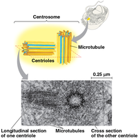
Centrosomes and Centrioles
Centrosomes and centrioles organize cell division in animal cells. The centrosome contains a pair of centrioles, which are composed of microtubules arranged in a specific pattern.
Cilia and Flagella
Flagella are long, whip-like extensions that propel cells through their environment. Cilia are shorter and more numerous, moving fluid across the cell surface or moving the cell itself.
Extracellular Components and Cell Connections
Cell Walls of Plants
The cell wall is an extracellular structure distinguishing plant cells from animal cells. It protects the cell, maintains its shape, and prevents excessive water uptake. Plant cell walls are made of cellulose fibers embedded in polysaccharides and protein.
Extracellular Matrix (ECM) of Animal Cells
Animal cells lack cell walls but have an elaborate extracellular matrix (ECM) composed of glycoproteins and other molecules. The ECM regulates cell behavior and gene activity through integrins and mechanical signaling.
Cell Junctions
Cells in tissues, organs, or organ systems adhere, interact, and communicate through direct physical contact. Three types of cell junctions are common in animal tissues:
Tight junctions: Prevent leakage of extracellular fluid
Desmosomes: Fasten cells together into strong sheets
Gap junctions: Provide cytoplasmic channels for communication
Plasmodesmata in Plant Cells
Plasmodesmata are channels connecting plant cells, allowing water, solutes, proteins, and RNA to pass from cell to cell.
Summary Table: Comparison of Cell Types and Structures
Feature | Prokaryotic Cells | Eukaryotic Cells |
|---|---|---|
Nucleus | No | Yes (double membrane) |
Membrane-bound organelles | No | Yes |
Cell size | Small | Larger |
Cell wall | Yes (most) | Plants (yes), Animals (no) |
DNA location | Nucleoid | Nucleus |
Additional info: The notes expand on brief points from the original slides, providing definitions, examples, and context for each cell structure and function. Images included are directly relevant to the explanation of cell compartmentalization and centrosome structure.
