 Back
BackBlood: Composition, Function, and Clinical Relevance
Study Guide - Smart Notes
 Tailored notes based on your materials, expanded with key definitions, examples, and context.
Blood: Composition and Function
Overview of Blood as a Connective Tissue
Blood is a specialized fluid connective tissue essential for transporting substances throughout the body. It consists of cells suspended in a liquid extracellular matrix called plasma. Blood links body systems by transporting oxygen, nutrients, hormones, and waste products.
Key Components: Plasma, erythrocytes (red blood cells), leukocytes (white blood cells), and thrombocytes (platelets).
Function: Transport, regulation, and protection.
Composition of Blood
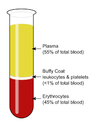
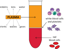
Plasma: The liquid matrix, making up about 55% of total blood volume. Contains water, proteins, ions, nutrients, wastes, and gases.
Buffy Coat: Thin layer containing leukocytes and platelets (<1% of blood volume).
Erythrocytes: Red blood cells, comprising about 45% of blood volume.
Plasma
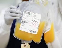
Plasma is a straw-colored fluid that serves as the medium for transporting nutrients, hormones, and waste products. It also plays a critical role in maintaining blood pH and osmotic balance.
Main Components: Water (90%), proteins, electrolytes, nutrients, hormones, and waste products.
Functions: Transport, buffering, and maintaining osmotic pressure.
Blood Proteins
Blood proteins are primarily found in plasma and are essential for maintaining osmotic potential, transporting substances, supporting the immune system, and enabling blood clotting.
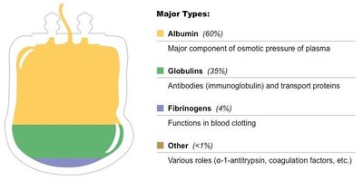
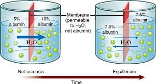
Albumin: Main protein responsible for osmotic pressure and transport of enzymes, vitamins, and hormones.
Globulins: Support immune function and transport lipids, hormones, and cholesterol.
Fibrinogen: Key protein in blood coagulation, forming fibrin clots.
Protein | Percentage | Main Function |
|---|---|---|
Albumin | 60% | Osmotic pressure, transport |
Globulins | 35% | Immunity, transport proteins |
Fibrinogen | 4% | Blood clotting |
Other | <1% | Various (e.g., coagulation factors) |
Osmotic Potential and Buffering
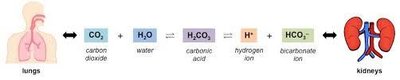
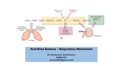
Albumin is crucial for regulating osmotic potential, which controls the movement of water between blood and tissues. Plasma also contains chemical buffers that maintain blood pH between 7.35 and 7.45, primarily through the carbonic acid-bicarbonate system.
Main Buffers: Carbonic acid (H2CO3) and bicarbonate ion (HCO3-).
Equation:
Blood Cells
Types of Blood Cells
Blood contains three main types of cells: erythrocytes, leukocytes, and thrombocytes. Each type has specialized functions essential for homeostasis.
Erythrocytes (Red Blood Cells): Transport oxygen and carbon dioxide.
Leukocytes (White Blood Cells): Defend against pathogens and foreign substances.
Thrombocytes (Platelets): Involved in blood clotting.
Erythrocytes (Red Blood Cells)
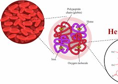
Erythrocytes are small, biconcave cells lacking a nucleus, mitochondria, and endoplasmic reticulum, maximizing space for hemoglobin. Their shape increases surface area for gas exchange.
Hemoglobin: Protein that binds and transports oxygen.
Function: Efficient oxygen delivery to tissues and removal of carbon dioxide.
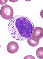
Leukocytes (White Blood Cells)
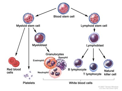
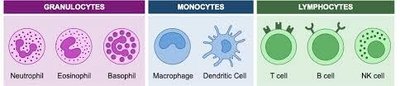
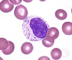
Leukocytes are part of the immune system and are classified into granulocytes (neutrophils, eosinophils, basophils), monocytes (macrophages), and lymphocytes (B cells, T cells, NK cells).
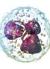
Granulocytes: Neutrophils are the most abundant, involved in phagocytosis of pathogens.
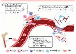
Monocytes: Differentiate into macrophages, which engulf and digest pathogens and debris.
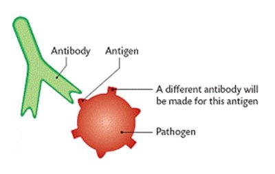
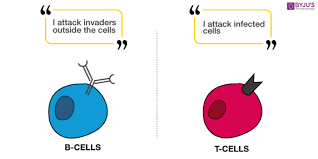
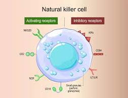
Lymphocytes: B cells produce antibodies; T cells destroy infected or cancerous cells; NK cells target virus-infected and tumor cells.
Thrombocytes (Platelets)
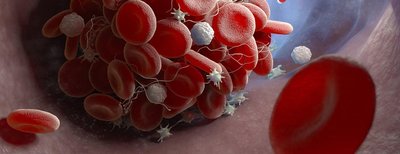
Platelets are small, colorless cell fragments produced in the bone marrow. They play a vital role in hemostasis by initiating blood clotting when blood vessels are injured.
Function: Adhere to damaged vessel walls, release clotting factors, and form a platelet plug.
Blood Clotting (Coagulation)
Mechanism of Blood Clotting
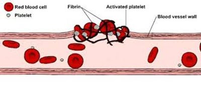
Blood clotting is a complex cascade that prevents excessive bleeding when vessels are injured. Platelets adhere to exposed collagen, and a series of enzymatic reactions convert prothrombin to thrombin, which then converts fibrinogen to fibrin, forming a stable clot.
Key Steps:
Platelet adhesion and activation
Release of thromboplastin
Conversion of prothrombin to thrombin
Formation of fibrin mesh
Clotting Rate and Clinical Testing
Clotting rates are measured to diagnose bleeding disorders and monitor anticoagulant therapy. The normal clotting time is 9–13 seconds. The International Normalised Ratio (INR) standardizes clotting time measurements for patients on blood thinners.
Clinical Relevance of Blood
Blood Tests and Diagnosis
Blood tests are essential for diagnosing various conditions. Abnormal values can indicate infection, clotting disorders, metabolic imbalances, or organ dysfunction.
High white blood cell count: Infection or inflammation
Fast/slow clotting rate: Clotting disorders
Elevated glucose: Diabetes
High cholesterol: Cardiovascular risk
High waste products: Kidney dysfunction
Bruising and Contusions
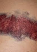
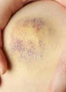
Bruising occurs when blood leaks into tissues due to vessel damage, causing discoloration. Contusions are deeper injuries affecting muscle or bone. The color changes as hemoglobin breaks down into biliverdin (green) and bilirubin (yellow), which is eventually processed by the liver.
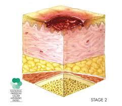
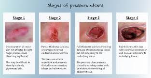
Pressure Sores (Bedsores)
Pressure sores are localized injuries to skin and underlying tissue due to prolonged pressure, often over bony areas. They result from restricted blood flow and can progress to tissue necrosis if untreated.
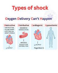
Physiological Shock
Physiological shock occurs when blood volume or pressure drops, leading to inadequate tissue perfusion and oxygen delivery. Symptoms include pale, cold skin, rapid breathing, weak pulse, dizziness, and nausea. Types of shock include hypovolemic, cardiogenic, distributive, and obstructive.
Additional info: This guide integrates foundational biology concepts with clinical applications, supporting topics such as cell components, the circulatory system, and immune system function.
