Back
BackCell Cycle Regulation, Cancer, and Chromosomal Mutations
Study Guide - Smart Notes
 Tailored notes based on your materials, expanded with key definitions, examples, and context.
Cell Cycle Regulation
Overview of Cell Cycle Control
The cell cycle is a tightly regulated process that ensures proper growth, development, and maintenance in multicellular organisms. The timing and rate of cell division vary among different cell types and are controlled by molecular signals within the cell.
Cell cycle regulation is crucial for normal tissue function.
Some cells, like skin cells, divide frequently, while others, such as nerve and muscle cells, rarely or never divide after maturity.
Regulation occurs at the molecular level, involving both internal and external signals.
Cytoplasmic Signals and Cell Cycle Control
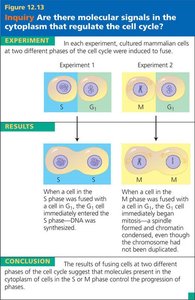
Experiments have shown that the cell cycle is driven by specific molecular signals present in the cytoplasm. These signals can trigger and coordinate key events in the cell cycle.
Fusing cells at different phases of the cell cycle demonstrates that cytoplasmic factors can induce progression through the cycle.
Factors Affecting Cell Division
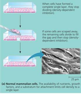
Anchorage dependence: Animal cells must be attached to a solid surface to divide.
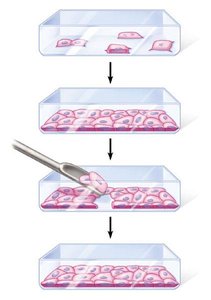
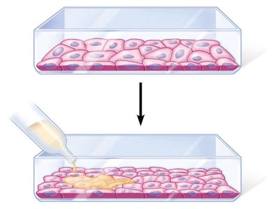
Density-dependent inhibition: Cells stop dividing when they form a single layer and touch each other. If cells are removed, neighboring cells divide to fill the gap.
Growth factors: Proteins secreted by cells that stimulate cell division. When growth factors are depleted, cell division stops.
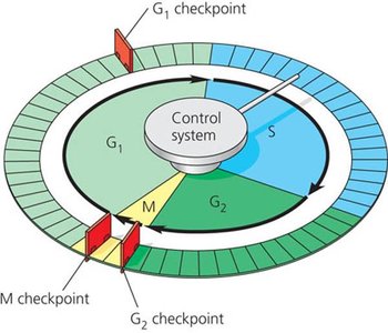
Cell Cycle Control System and Checkpoints
The cell cycle control system is a set of molecules that operate cyclically to trigger and coordinate cell cycle events. The system can proceed on its own but is regulated at checkpoints by internal and external controls.
Checkpoints: Critical control points where stop and go-ahead signals regulate the cycle.
Three major checkpoints: G1, G2, and M phases.

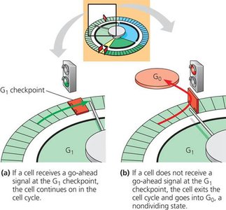
G1 Checkpoint and the G0 Phase
The G1 checkpoint is the most crucial for mammalian cells. If a cell receives a go-ahead signal at G1, it will complete the cycle and divide. If not, it enters a nondividing state called the G0 phase.
Most human body cells are in the G0 phase.
Some cells, like liver cells, can re-enter the cycle if stimulated by growth factors.
Internal and External Regulation at Checkpoints
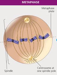
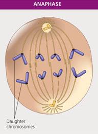
Internal signals: For example, during the M phase checkpoint, anaphase does not begin until all chromosomes are properly attached to the spindle at the metaphase plate. This ensures accurate chromosome separation.
External signals: Factors such as nutrients and growth factors in the environment can influence cell division.
Loss of Cell Cycle Control and Cancer
Characteristics of Cancer Cells
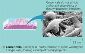
Cancer cells do not respond to normal cell cycle controls. They may divide uncontrollably, ignore density-dependent inhibition, and continue dividing even when crowded.
Cancer cells can synthesize their own growth factors or divide without them.
They may stop dividing at random points in the cycle, not at checkpoints.
In culture, cancer cells can divide indefinitely (immortal), unlike normal cells.
Formation and Spread of Cancer
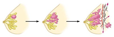
Transformation: A single cell undergoes genetic changes and becomes cancerous.
Benign tumor: Abnormal cells remain at the original site and can often be removed surgically.
Malignant tumor: Cells invade neighboring tissues and can impair organ function. The spread of cancer cells to distant sites is called metastasis.
Treatment of Cancer
Localized tumors may be treated with radiation, which damages cancer cell DNA.
Metastatic tumors are treated with chemotherapy, which targets dividing cells but also affects normal cells, causing side effects.
Mutations and Chromosomal Abnormalities
What Are Mutations?
Mutations are changes in the nucleotide sequence of DNA. They can occur in somatic cells (not inherited) or gametes (inherited). Most mutations are neutral, but some can be harmful or beneficial.
Caused by errors in DNA replication, chemicals, or radiation.
Some mutations are repaired by enzymes.
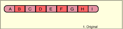
Types of Chromosome Mutations
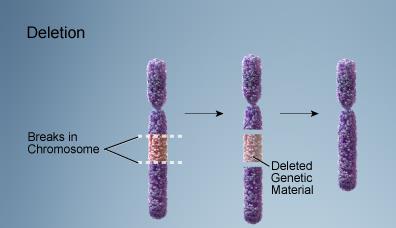
Deletion: Loss of a chromosome segment.
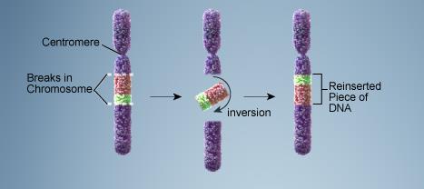
Inversion: A chromosome segment breaks off, flips, and reattaches.
Duplication: A segment is repeated.
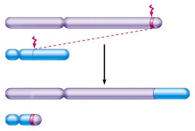
Translocation: A segment moves to a non-homologous chromosome.
Nondisjunction: Failure of chromosomes to separate during meiosis, leading to abnormal chromosome numbers.

Gene Mutations
Point mutations: Change in a single nucleotide (substitution, insertion, or deletion).
Frameshift mutations: Insertion or deletion of nucleotides that alters the reading frame, leading to incorrect protein synthesis.
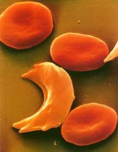
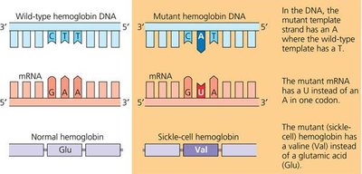
Example: Sickle cell disease is caused by a single nucleotide substitution in the hemoglobin gene.
Chromosomal Syndromes and Nondisjunction
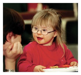
Nondisjunction during meiosis can result in gametes with abnormal chromosome numbers, leading to syndromes such as Down syndrome (trisomy 21), Klinefelter syndrome (XXY), and Turner syndrome (XO).
Down syndrome: Caused by an extra copy of chromosome 21. Incidence increases with maternal age.
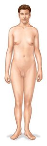
Klinefelter syndrome: Males with an extra X chromosome (XXY).
Turner syndrome: Females with only one X chromosome (XO).
Chromosomal Translocations and Cancer
Translocations in somatic cells can lead to cancers such as chronic myelogenous leukemia (CML), where a reciprocal translocation between chromosomes 9 and 22 creates the "Philadelphia chromosome." This activates a cancer-causing gene.
Cell Division in Bacteria
Binary Fission in Prokaryotes
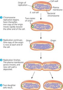
Bacteria reproduce by binary fission, a process in which the cell divides in half to produce two genetically identical daughter cells. The bacterial chromosome is circular and replication begins at a specific origin.
Replication starts at the origin and proceeds around the chromosome.
As replication continues, the cell elongates and the origins move to opposite ends.
When replication is complete, the cell divides, and each daughter cell receives a complete genome.