 Back
BackCell Division, Cell Cycle, and Genetic Stability: A Comprehensive Study Guide
Study Guide - Smart Notes
 Tailored notes based on your materials, expanded with key definitions, examples, and context.
Cell Division and Reproduction
Asexual Reproduction
Asexual reproduction is a mode of reproduction that does not involve the fusion of gametes. Offspring are genetically identical to the parent, as they inherit the same DNA. This process is common in unicellular organisms and some multicellular organisms.
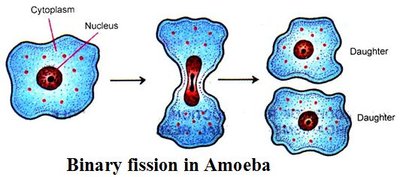
Binary Fission: A form of asexual reproduction in prokaryotes and some single-celled eukaryotes, where the cell divides into two genetically identical daughter cells.
Advantages: Rapid population growth, no need for a mate, offspring are well-adapted to stable environments.
Disadvantages: Lack of genetic diversity, which can be detrimental in changing environments.
Example: Amoeba reproducing by binary fission.
Sexual Reproduction
Sexual reproduction involves the production and fusion of gametes (sperm and egg), resulting in genetically unique offspring. This process increases genetic diversity and is essential for evolution and adaptation.
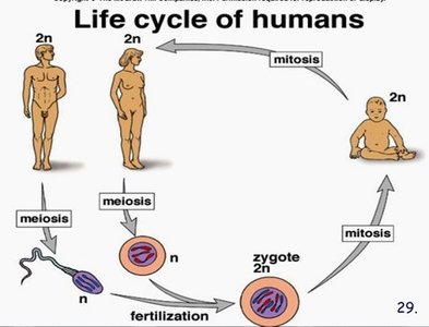
Meiosis: Specialized cell division that reduces chromosome number by half, producing haploid gametes.
Fertilization: Fusion of two gametes to form a diploid zygote.
Advantages: Genetic variation, increased adaptability.
Disadvantages: Requires more energy and time, finding a mate can be challenging.
Example: Human life cycle involving meiosis, fertilization, and mitosis.
Genetic Material: Prokaryotes vs. Eukaryotes
Prokaryotic DNA
Prokaryotes (bacteria and archaea) have a simpler organization of genetic material compared to eukaryotes.
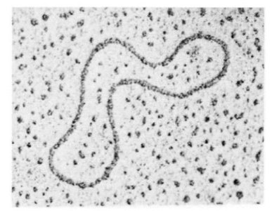
Location: DNA is found freely in the cytoplasm.
Structure: Circular DNA, not associated with histone proteins (naked DNA).
Plasmids: Small, extrachromosomal DNA molecules.
Genome: Compact, with little repetitive DNA.
Eukaryotic DNA
Eukaryotes (plants, animals, fungi, protists) have more complex genetic material.
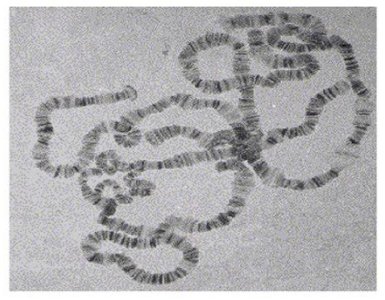
Location: DNA is contained within a nucleus.
Structure: Linear chromosomes, DNA is wrapped around histone proteins to form chromatin.
Genome: Contains large amounts of non-coding and repetitive DNA.
Organelles: Mitochondria and chloroplasts may have their own DNA.
Viruses and Their Reproduction
Types of Viruses
Viruses can infect both prokaryotic and eukaryotic cells, but their genetic material and reproductive strategies differ.
Prokaryotic Viruses (Bacteriophages): Infect bacteria, less diverse in genome type.
Eukaryotic Viruses: Infect eukaryotic cells, more diverse (dsDNA, ssDNA, dsRNA, ssRNA, etc.).
Lytic vs. Lysogenic Cycles
Viruses can reproduce via two main cycles:
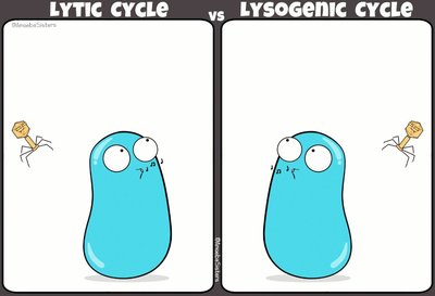
Lytic Cycle: Virus replicates inside the host, causing cell lysis and release of new viruses.
Lysogenic Cycle: Viral DNA integrates into the host genome and remains dormant until triggered to enter the lytic cycle.
Chromosome Structure and Karyotypes
Chromosome, Chromatid, and Chromatin
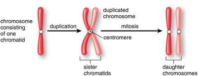
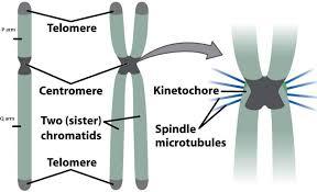
Chromosomes are highly organized structures of DNA and proteins. During cell division, DNA condenses into visible chromosomes.
Chromosome: A single, long DNA molecule with associated proteins.
Chromatid: Each of the two identical halves of a duplicated chromosome.
Centromere: Region where sister chromatids are joined.
Chromatin: The less condensed form of DNA present during interphase.
Diploid vs. Haploid Cells
Organisms can have cells with different chromosome numbers:
Diploid (2n): Two sets of chromosomes (somatic cells).
Haploid (n): One set of chromosomes (gametes).
Example: Humans have 46 chromosomes in somatic cells (2n = 46) and 23 in gametes (n = 23).
Karyotypes
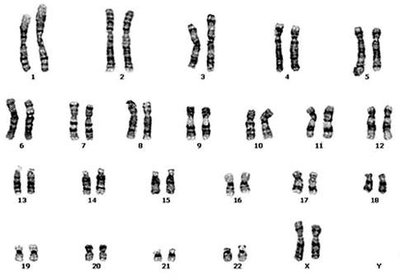
A karyotype is an organized profile of an individual's chromosomes, used to detect chromosomal abnormalities and determine sex.
Autosomes: Non-sex chromosomes.
Allosomes: Sex chromosomes (X and Y).
Applications: Diagnosis of genetic disorders, determination of sex.
The Cell Cycle and Mitosis
Phases of the Cell Cycle
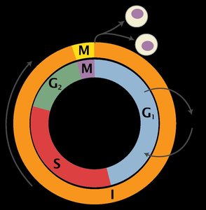
The cell cycle is the series of events that cells go through as they grow and divide. It consists of interphase, mitosis, and cytokinesis.
Interphase: Includes G1 (growth), S (DNA synthesis), and G2 (preparation for mitosis).
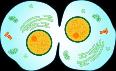
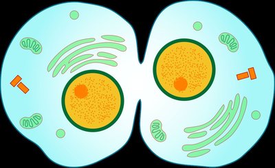
Mitosis (M phase): Division of the nucleus.
Cytokinesis: Division of the cytoplasm.
Phases of Mitosis
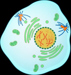
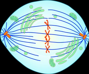
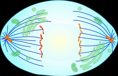
Mitosis is divided into four main stages, followed by cytokinesis:
Prophase: Chromosomes condense, nuclear envelope dissolves, spindle fibers form.
Metaphase: Chromosomes align at the cell's equator.
Anaphase: Sister chromatids are pulled apart to opposite poles.
Telophase: Nuclear envelope reforms, chromosomes decondense.
Cytokinesis: Cytoplasm divides, forming two identical daughter cells.
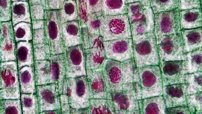
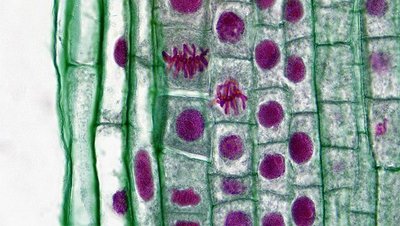
Microscopic Observation of Mitosis
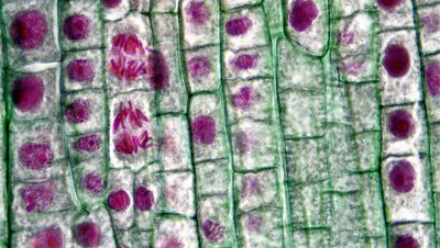
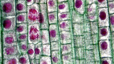
Onion root tip cells are commonly used to observe the stages of mitosis under a microscope.
Cell Cycle Regulation and Cancer
Cell Cycle Checkpoints
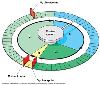
The cell cycle is regulated by checkpoints at G1, G2, and M phases to ensure proper division and prevent errors.
G1 Checkpoint: Checks for cell size, nutrients, and DNA damage.
G2 Checkpoint: Ensures DNA replication is complete and checks for DNA damage.
M Checkpoint: Ensures all chromosomes are properly attached to the spindle before anaphase.
Fate of Cells: Division, Differentiation, and Apoptosis
Cells can divide, differentiate into specialized types, or undergo programmed cell death (apoptosis) to maintain tissue health and function.
Cell Division: Produces new cells for growth and repair.
Differentiation: Cells become specialized for specific functions.
Apoptosis: Programmed cell death removes damaged or unnecessary cells.
Cancer and Uncontrolled Cell Division
Cancer results from the loss of cell cycle control, often due to mutations in genes that regulate division. Tumors can be benign (localized) or malignant (invasive and metastatic).
Causes: DNA damage from carcinogens, radiation, viruses, or inherited mutations.
Treatments: Surgery, radiation, chemotherapy.
Angiogenesis: Tumors stimulate new blood vessel growth to supply nutrients.
Meiosis and Genetic Variation
Overview of Meiosis
Meiosis is a two-part cell division process that produces four genetically unique haploid gametes from a diploid parent cell. It is essential for sexual reproduction and genetic diversity.
Meiosis I: Homologous chromosomes separate.
Meiosis II: Sister chromatids separate.
Key Events: Crossing over (genetic recombination), independent assortment.
Gametogenesis
Formation of gametes differs between males (spermatogenesis) and females (oogenesis).
Spermatogenesis: Produces four haploid sperm cells from each diploid spermatogonium.
Oogenesis: Produces one mature ovum and polar bodies from each diploid oocyte.
Chromosomal Mutations and Nondisjunction
Types of Chromosomal Mutations
Chromosomal mutations can occur during crossing over in meiosis or due to errors in DNA repair. These mutations can have severe effects if they occur in gametes.
Mutation Type | Description |
|---|---|
Deletion | An entire section of a chromosome is missing. |
Duplication | One section of the chromosome is doubled. |
Inversion | A section of a chromosome has its gene sequences reversed. |
Insertion | Part of one chromosome is inserted into a different chromosome. |
Translocation | Two non-homologous chromosomes exchange alleles during crossing over. |
Nondisjunction and Aneuploidy
Nondisjunction is the failure of chromosomes to separate properly during meiosis, resulting in gametes with abnormal chromosome numbers (aneuploidy).
Meiosis I Nondisjunction: All daughter cells are aneuploid.
Meiosis II Nondisjunction: Half of the daughter cells are aneuploid.
Examples: Down syndrome (trisomy 21), Klinefelter syndrome (XXY), Turner syndrome (X0).
Summary Table: Chromosomal Mutation Types
Mutation Type | Description |
|---|---|
Deletion | An entire section of a chromosome is missing. |
Duplication | One section of the chromosome is doubled. |
Inversion | A section of a chromosome has its gene sequences reversed. |
Insertion | Part of one chromosome is inserted into a different chromosome. |
Translocation | Two non-homologous chromosomes exchange alleles during crossing over. |
Key Equations and Concepts
Diploid Number:
Haploid Number:
DNA Replication:
Chromosome Number in Humans: ,
