 Back
BackUnit 4
Study Guide - Smart Notes
 Tailored notes based on your materials, expanded with key definitions, examples, and context.
Cell Division and Reproduction
Sexual vs. Asexual Reproduction
Reproduction is the biological process by which new individual organisms are produced. It can occur via two main mechanisms: sexual and asexual reproduction.
Asexual reproduction: Involves a single parent and produces genetically identical offspring (clones). Common in unicellular organisms and some plants and animals.
Sexual reproduction: Involves two parents and the fusion of gametes, resulting in genetically diverse offspring.
Examples of Asexual Reproduction
Budding in hydra
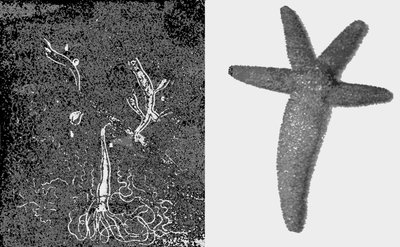
Regeneration in starfish
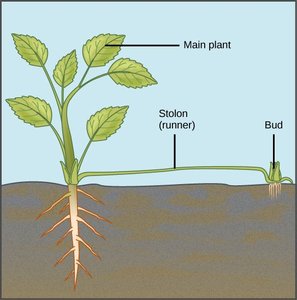
Vegetative propagation in plants (e.g., runners/stolons in strawberries)
Advantages of Asexual Reproduction
Rapid population increase
No need for a mate
Preservation of successful genotypes in stable environments
Types of Cell Division
Binary Fission, Mitosis, and Meiosis
Cell division is essential for growth, development, and reproduction. There are three main types:
Type | Process | Result | Purpose |
|---|---|---|---|
Binary Fission | DNA replication; cell splits in two | Two identical cells | Asexual reproduction (prokaryotes) |
Mitosis | DNA replication; segregation; cell splits | Two identical daughter cells | Growth, development, tissue repair (eukaryotes) |
Meiosis | DNA replication; two rounds of division | Four haploid gametes | Sexual reproduction (eukaryotes) |
The Cell Cycle and Mitosis
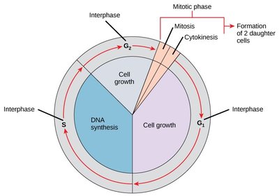
The Cell Cycle
The cell cycle is the ordered sequence of events that a cell goes through between one division and the next. It consists of interphase (G1, S, G2) and the mitotic phase (mitosis and cytokinesis).
G1 phase: Cell growth
S phase: DNA synthesis (replication)
G2 phase: Preparation for mitosis
Mitotic phase: Mitosis (nuclear division) and cytokinesis (cytoplasmic division)
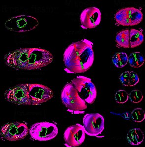
Mitosis: An Overview
Mitosis is the process by which a eukaryotic cell separates its duplicated chromosomes into two identical sets, resulting in two genetically identical daughter cells.
Prophase: Chromosomes condense, spindle forms
Metaphase: Chromosomes align at the cell equator
Anaphase: Sister chromatids separate
Telophase: Nuclear envelopes reform
Cytokinesis: Division of the cytoplasm

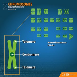
Chromosomes and Chromatin
Chromosomes are structures made of DNA and proteins (chromatin) that carry genetic information. In humans, there are 46 chromosomes (23 pairs).
Chromatin: DNA-protein complex that is diffuse in non-dividing cells
Chromosome: Condensed form of chromatin visible during cell division
Gene: Segment of DNA encoding a trait
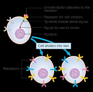
Cell Cycle Regulation and Cancer
Cell Cycle Checkpoints
Cell cycle checkpoints ensure that cells only proceed to the next stage when conditions are favorable and DNA is undamaged.
G1 checkpoint: Checks for cell size, nutrients, growth factors, and DNA damage
G2 checkpoint: Checks for DNA replication completion and damage
M checkpoint: Ensures chromosomes are properly attached to the spindle
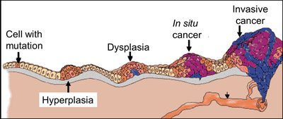
Cancer and the Cell Cycle
Cancer results from uncontrolled cell division due to failures in cell cycle regulation. Tumors can be benign (localized) or malignant (capable of metastasis).
Transformation: Process by which a normal cell becomes cancerous
Metastasis: Spread of cancer cells to other parts of the body
Cancer therapies: Surgery, radiation, chemotherapy (targets dividing cells)
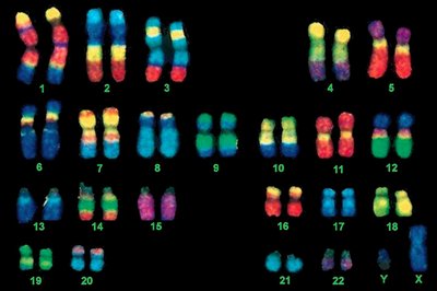
Chromosome Number and Structure
Diploid and Haploid Cells
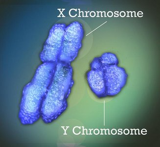
Organisms have two sets of chromosomes (diploid, 2n) in somatic cells and one set (haploid, n) in gametes. Humans have 23 pairs of chromosomes: 22 pairs of autosomes and 1 pair of sex chromosomes (XX or XY).
Meiosis and Genetic Diversity
Overview of Meiosis
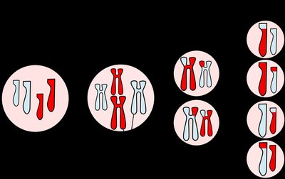
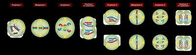
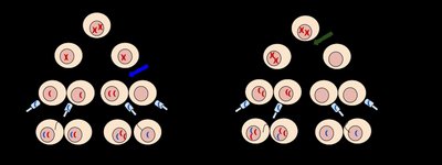
Meiosis is a specialized form of cell division that reduces the chromosome number by half, producing four genetically unique haploid gametes. It consists of two sequential divisions: meiosis I and meiosis II.
Meiosis I: Homologous chromosomes separate
Meiosis II: Sister chromatids separate
Sources of Genetic Variation
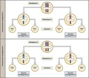
Independent assortment: Random alignment of homologous chromosomes during metaphase I
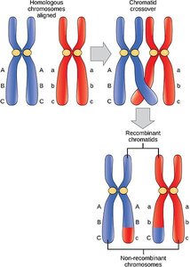
Crossing over: Exchange of genetic material between non-sister chromatids during prophase I
Random fertilization: Any sperm can fertilize any egg
Nondisjunction and Aneuploidy
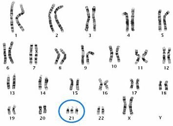
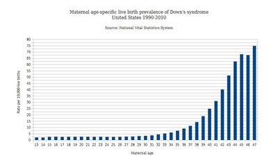
Nondisjunction is the failure of chromosomes to separate properly during meiosis, leading to gametes with abnormal chromosome numbers (aneuploidy).
Down syndrome: Trisomy 21 (three copies of chromosome 21)
Turner syndrome: XO (single X chromosome)
Klinefelter syndrome: XXY (extra X chromosome in males)
Mendelian Genetics
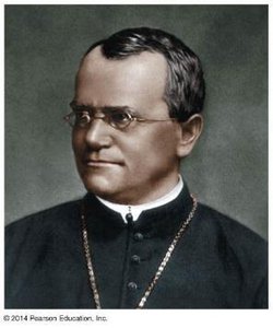
Gregor Mendel and the Laws of Inheritance
Gregor Mendel's experiments with pea plants established the basic principles of heredity, including the law of segregation and the law of independent assortment.
Law of Segregation: Each individual has two alleles for each gene, which segregate during gamete formation.
Law of Independent Assortment: Genes for different traits assort independently during gamete formation.
Monohybrid Crosses and Punnett Squares
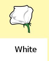
Punnett squares are used to predict the genotypic and phenotypic ratios of offspring from genetic crosses.
Genotype: Genetic makeup (e.g., PP, Pp, pp)
Phenotype: Observable trait (e.g., purple or white flowers)
Dihybrid Crosses and Probability
Dihybrid crosses involve two traits and demonstrate independent assortment. Probability rules (product and addition) are used to calculate genotype and phenotype ratios.
Extensions and Exceptions to Mendel's Laws
Incomplete dominance: Heterozygotes show an intermediate phenotype (e.g., pink flowers from red and white parents)
Codominance: Both alleles are expressed (e.g., ABO blood groups)
Polygenic inheritance: Traits influenced by multiple genes (e.g., height, skin color)
Linkage: Genes located close together on the same chromosome tend to be inherited together
DNA: The Genetic Material
Structure and Function of DNA
DNA (deoxyribonucleic acid) is the hereditary material in all living organisms. It encodes genetic information using four nucleotide bases: adenine (A), thymine (T), cytosine (C), and guanine (G).
Double helix: Two strands held together by complementary base pairing (A-T, C-G)
Central Dogma: DNA → RNA → Protein
Gene Expression
Transcription: Synthesis of RNA from a DNA template
Translation: Synthesis of protein from an mRNA template at the ribosome
Mutations
Mutations are changes in the DNA sequence that can affect gene function. Types include:
Substitution: One base is replaced by another
Insertion/Deletion (indels): Addition or loss of bases, potentially causing frameshifts
Example: Sickle-cell anemia is caused by a single nucleotide substitution in the hemoglobin gene.
Summary Table: Types of Cell Division
Type | Organisms | Purpose | Result |
|---|---|---|---|
Binary Fission | Bacteria, Archaea | Asexual reproduction | 2 identical cells |
Mitosis | Eukaryotes | Growth, repair, asexual reproduction | 2 identical cells |
Meiosis | Eukaryotes | Sexual reproduction | 4 genetically unique gametes |
