 Back
BackLEC 6: Cell Membranes, Transport, and Cell Walls: Structure and Function
Study Guide - Smart Notes
 Tailored notes based on your materials, expanded with key definitions, examples, and context.
Bulk Transport Across Membranes
Exocytosis and Endocytosis
Cells use bulk transport mechanisms to move large molecules or particles that cannot pass through transporter proteins. These processes require energy and involve the formation of vesicles from the flexible plasma membrane.
Exocytosis: The process by which cells export substances (e.g., proteins, polysaccharides) out of the cell via secretory vesicles that fuse with the plasma membrane.
Endocytosis: The process of importing substances into the cell by engulfing them in vesicles. There are three main types:
Phagocytosis: "Cell eating"; the cell engulfs large particles or even other cells.
Pinocytosis: "Cell drinking"; the cell takes in extracellular fluid and dissolved solutes.
Receptor-mediated endocytosis: Specific molecules (ligands) bind to receptors on the cell surface, triggering vesicle formation for selective uptake.
Example: Uptake of cholesterol by cells via LDL (low-density lipoprotein) particles is mediated by receptor-mediated endocytosis.
Receptor-Mediated Endocytosis and Clathrin
Receptor-mediated endocytosis involves the binding of specific ligands to cell surface receptors, which cluster in regions of the membrane coated by the protein clathrin. Clathrin helps form a "pit" that buds off into a vesicle, allowing the cell to internalize the ligand. After vesicle formation, clathrin is released and receptors can be recycled.
Clathrin: A coat protein with a triskelion shape that facilitates vesicle formation.
Ligand: A molecule that specifically binds to a receptor.
Lipoproteins and Cholesterol Transport
Lipoproteins are complexes that transport fats through the bloodstream. Cells recognize specific apolipoproteins on the surface of lipoproteins, such as Apolipoprotein B-100 on LDL, and internalize them via receptor-mediated endocytosis.
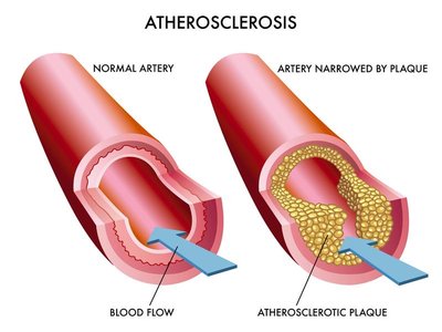
LDL (Low-Density Lipoprotein): Delivers cholesterol to cells; high levels are associated with increased risk of atherosclerosis.
HDL (High-Density Lipoprotein): Removes excess cholesterol from tissues and transports it to the liver for excretion; considered "good" cholesterol.
Clinical Relevance: High LDL and low HDL levels are risk factors for cardiovascular disease due to cholesterol buildup in arteries (atherosclerosis).
Transporter Proteins and Membrane Transport
Types of Transporters
Transport proteins facilitate the movement of molecules across membranes. They are classified based on their mechanism and directionality:
Uniporters: Transport a single type of molecule in one direction.
Symporters: Transport two different molecules in the same direction.
Antiporters: Transport two different molecules in opposite directions.
Transport can be passive (facilitated diffusion) or active:
Primary Active Transport: Uses ATP directly (e.g., Na+/K+ pump).
Secondary Active Transport: Uses ion gradients established by primary active transport to drive movement of other molecules.
Review of Membrane Structure and Function
Cell membranes are fluid mosaics of lipids and proteins, allowing selective permeability.
Passive transport moves substances down their concentration gradients without energy input.
Active transport moves substances against gradients, requiring energy.
Bulk transport (exocytosis/endocytosis) moves large particles or volumes.
Cell Walls: Structure and Function
Bacterial Cell Walls
Bacterial cell walls provide shape and protect cells from osmotic lysis. Most bacterial cell walls contain peptidoglycan, a polymer unique to bacteria.
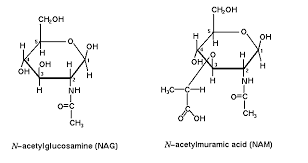
Peptidoglycan: Composed of repeating units of N-acetylglucosamine (NAG) and N-acetylmuramic acid (NAM), cross-linked by short peptides.
Gram Stain: Differentiates bacteria based on cell wall structure:
Gram-positive: Thick peptidoglycan layer, stains purple.
Gram-negative: Thin peptidoglycan layer plus outer membrane, stains pink/red.
Peptidoglycan Synthesis: Occurs only in growing cells and is not a barrier to solutes. Cross-linking (transpeptidation) adds strength to the cell wall.
Antibiotics Targeting Cell Walls
Some antibiotics target bacterial cell walls, exploiting differences between prokaryotic and eukaryotic cells.
Lysozyme: An enzyme in bodily fluids that hydrolyzes β 1-4 linkages between NAG and NAM, leading to cell lysis.
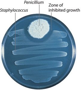
Penicillin: Inhibits the enzyme responsible for transpeptidation, preventing cross-linking of peptidoglycan and causing cell lysis, especially in Gram-positive bacteria.
External Structures of Prokaryotes
Capsules and Pili
Capsule: A polysaccharide layer outside the cell wall that protects bacteria and aids in attachment to surfaces; rare in Archaea.
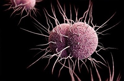
Pili: Hair-like appendages; fimbriae attach to surfaces, while sex pili facilitate DNA transfer (conjugation).
Motility: Flagella and Chemotaxis
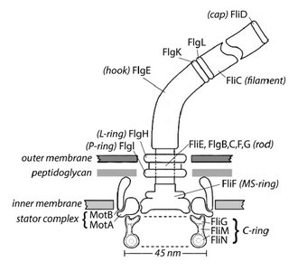
Many bacteria move using flagella, which are structurally and evolutionarily distinct from those in Archaea and Eukaryotes. Bacterial flagella rotate, powered by a proton (H+) or sodium (Na+) gradient, enabling "runs" and "tumbles" for chemotaxis (movement toward or away from chemical stimuli).
Cell Walls in Archaea and Eukaryotes
Archaeal Cell Walls
No peptidoglycan; may have related molecules or unique lipopolysaccharides.
Membranes contain ether-linked lipids, providing extra strength and stability.
Eukaryotic Cell Walls
Plants: Cell walls are composed of cellulose embedded in a matrix of other polysaccharides and proteins. The primary cell wall is thin and flexible; some cells add a secondary wall for extra strength (e.g., wood contains lignin).
Middle Lamella: The layer between adjacent plant cells, rich in pectins, glues cells together.
Plasmodesmata: Channels that connect plant cells, allowing passage of water and small solutes.
Fungi: Cell walls contain chitin.
Animals: No cell wall; instead, cells secrete an extracellular matrix (ECM) composed mainly of glycoproteins (e.g., collagen) and proteoglycans.

Cell Junctions in Animal Cells
Types of Cell Junctions
Tight Junctions: Seal neighboring cells together, preventing leakage of extracellular fluid (e.g., intestinal lining).
Desmosomes: Anchor cells together with strong protein filaments, providing mechanical strength (e.g., in muscle tissue).
Gap Junctions: Channels that allow ions and small molecules to pass directly between adjacent cells, enabling coordinated responses (e.g., in heart muscle).
Junction Type | Main Function | Example Tissue |
|---|---|---|
Tight Junction | Seals cells, prevents leakage | Intestinal epithelium |
Desmosome | Anchors cells, resists stress | Skin, muscle |
Gap Junction | Communication, ion flow | Heart muscle |
Additional info: The ECM also plays a role in cell signaling and tissue organization. Integrins are transmembrane proteins that connect the ECM to the cytoskeleton, transmitting signals and mechanical forces.
