 Back
BackCell Structure, Division, Genetics, and Evolution: Core Concepts for College Biology
Study Guide - Smart Notes
 Tailored notes based on your materials, expanded with key definitions, examples, and context.
Cell Structure and Microscopy
Microscopy and Cell Types
Microscopy is essential for studying cells, the basic units of life. Light microscopes use visible light to magnify specimens, while electron microscopes provide much higher resolution, allowing visualization of much smaller structures.
Magnification: The process of enlarging the appearance of an object.
Resolution: The ability to distinguish two close objects as separate entities.
Electron Microscopes: Can resolve structures as small as 200 nanometers, using electrons that bounce off surfaces.
Domains of Life
All living organisms are classified into three domains based on cellular structure:
Bacteria: Prokaryotic, single-celled organisms.
Archaea: Prokaryotic, but genetically distinct from bacteria.
Eukaryota: Organisms with membrane-bound organelles, including protists, plants, fungi, and animals.
Prokaryotic vs. Eukaryotic Cells
Cells are surrounded by a plasma membrane, contain chromosomes (genetic material), and ribosomes (for protein synthesis). Key differences include:
Prokaryotes: No membrane-bound nucleus; DNA is in a nucleoid region. Lack specialized organelles. Most have a cell wall for protection and structure.
Eukaryotes: Have a true nucleus enclosed by a nuclear envelope and various organelles. Generally larger (10–100 μm).
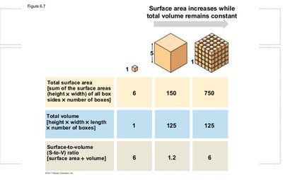
Surface Area to Volume Ratio
The surface area-to-volume ratio is crucial for cell function, affecting the rate of exchange of materials with the environment. Smaller cells have a higher ratio, facilitating efficient exchange.
Cell Structure and Organelles
Animal and Plant Cells
Animal Cells: Surrounded by a plasma membrane (phospholipid bilayer), which acts as a selective barrier.
Plant Cells: Have a rigid cell wall outside the plasma membrane for additional support.
Major Organelles
Cytoskeleton: Maintains cell shape and enables movement.
Endoplasmic Reticulum (ER): Rough ER (with ribosomes) synthesizes proteins; Smooth ER (without ribosomes) synthesizes lipids.
Nucleus: Contains genetic material; surrounded by a double membrane (nuclear envelope).
Golgi Apparatus: Modifies, sorts, and packages proteins and lipids.
Ribosomes: Sites of protein synthesis; can be free in cytosol or bound to ER.
Mitochondria: Produce ATP through cellular respiration; have inner folds called cristae.
Lysosomes: Contain hydrolytic enzymes for digestion of macromolecules.
Cell Division: Mitosis and Meiosis
Somatic vs. Germline Cells
Somatic Cells: Non-reproductive, diploid (2n), divide by mitosis.
Germline Cells: Reproductive, haploid (n), divide by meiosis; mutations can be inherited.
The Cell Cycle
The cell cycle consists of interphase (G1, S, G2) and the mitotic phase (mitosis and cytokinesis). Checkpoints ensure accurate division.
G1: Cell growth and energy accumulation.
S: DNA replication; chromosomes become sister chromatids.
G2: Preparation for mitosis; protein synthesis.
Mitosis: Division of the nucleus, followed by cytokinesis (division of cytoplasm).
G0: Quiescent phase; cells not actively dividing.
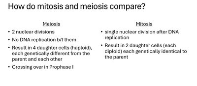
Mitosis vs. Meiosis
Mitosis and meiosis are two types of cell division with distinct outcomes.
Mitosis: One division, produces two genetically identical diploid cells.
Meiosis: Two divisions, produces four genetically unique haploid cells; includes crossing over in Prophase I.
Phases of Mitosis
Prophase: Chromosomes condense, spindle forms.
Prometaphase: Nuclear envelope fragments, spindle attaches to kinetochores.
Metaphase: Chromosomes align at the cell equator.
Anaphase: Sister chromatids separate to opposite poles.
Telophase: Nuclear envelopes reform, chromosomes decondense.
Cytokinesis: Division of cytoplasm (cleavage furrow in animals, cell plate in plants).
Phases of Meiosis
Meiosis I: Homologous chromosomes separate (crossing over occurs in Prophase I).
Meiosis II: Sister chromatids separate, similar to mitosis.
Genetics: Mendelian Principles
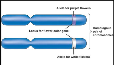
Genes, Alleles, and Chromosomes
Genes are units of heredity located on chromosomes. Alleles are different versions of a gene. Each individual inherits two alleles for each gene, one from each parent.
Mendel's Laws
Law of Segregation: Two alleles for a gene separate during gamete formation.
Law of Independent Assortment: Genes on different chromosomes are inherited independently.
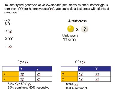
Test Cross and Punnett Squares
A test cross determines the genotype of an individual with a dominant phenotype by crossing with a homozygous recessive individual.
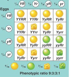
Dihybrid Cross and Phenotypic Ratios
Dihybrid crosses involve two genes and demonstrate independent assortment. The classic phenotypic ratio is 9:3:3:1 for two heterozygotes.
Extensions of Mendelian Genetics
Incomplete Dominance: Heterozygote shows intermediate phenotype.
Codominance: Both alleles are expressed equally.
Pleiotropy: One gene affects multiple traits (e.g., sickle cell disease).
Epistasis: One gene affects the expression of another gene.
Polygenic Inheritance: Multiple genes contribute to a single trait (e.g., skin color).
Lethal Alleles: Cause death when present in certain genotypes.
Sex-linked Traits: Genes located on sex chromosomes (e.g., hemophilia, color blindness).
Evolution and Population Genetics
Darwin's Theory of Evolution
Charles Darwin proposed that evolution occurs by natural selection, where individuals with advantageous traits survive and reproduce more successfully.
Evidence: Fossils, biogeography, direct observation, homology.
Key Concepts: Variation, inheritance, selection, and time drive evolutionary change.
Microevolution and Gene Pools
Microevolution refers to changes in allele frequencies within a population over time. The gene pool is the total collection of alleles in a population.
Hardy-Weinberg Equilibrium
The Hardy-Weinberg theorem describes a non-evolving population. Allele and genotype frequencies remain constant if five conditions are met: large population, no gene flow, no mutations, random mating, and no natural selection.
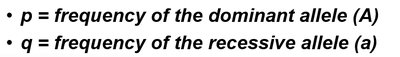
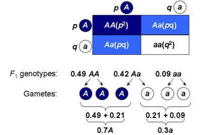
Allele Frequencies: p = frequency of dominant allele (A); q = frequency of recessive allele (a).
The Hardy-Weinberg equation is:
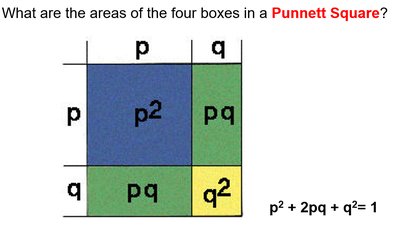
p2: Frequency of homozygous dominant genotype (AA)
2pq: Frequency of heterozygous genotype (Aa)
q2: Frequency of homozygous recessive genotype (aa)
Example: If p = 0.7 and q = 0.3, then:
, ,
Summary Table: Mitosis vs. Meiosis
Meiosis | Mitosis | |
|---|---|---|
Number of Divisions | 2 | 1 |
DNA Replication Between Divisions | No | Yes (before division) |
Number of Daughter Cells | 4 (haploid, genetically unique) | 2 (diploid, genetically identical) |
Crossing Over | Yes (Prophase I) | No |
