 Back
BackCell Transport and the Cell Cycle: Passive and Active Transport, Mitosis, and Meiosis
Study Guide - Smart Notes
 Tailored notes based on your materials, expanded with key definitions, examples, and context.
Cell Transport Mechanisms
Passive and Active Transport
Cells use various mechanisms to move substances across their membranes, maintaining homeostasis and supporting cellular functions.
Passive Transport: Movement of molecules from high to low concentration, following the concentration gradient. No energy (ATP) is required. Examples include gas exchange in lungs and capillaries.
Active Transport: Movement of molecules against the concentration gradient (low to high concentration), requiring energy in the form of ATP. Occurs in nerves, kidney re-absorption, and absorption of glucose and amino acids in the intestines.
Osmosis: The diffusion of water molecules from an area of low solute concentration to high solute concentration, crucial in kidney function and across all cell membranes.
Facilitated Diffusion: Passive transport of molecules through protein channels, important in neurons and muscle cells.
Process | Energy Required? | Direction | In the Body |
|---|---|---|---|
Passive Transport | No | High → Low | Lungs, capillaries, kidneys |
Active Transport | Yes (ATP) | Low → High | Neurons, kidneys, intestines |
Osmosis | No | Water: Low → High solute | Kidneys, all cells |
Facilitated Diffusion | No | High → Low | Muscles, RBCs, neurons |
The Cell Cycle
Overview and Importance
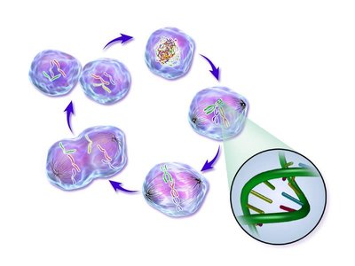
The cell cycle is the series of stages that cells undergo to grow and divide, ensuring tissue growth, repair, and replacement of old cells. It consists of Interphase and Cell Division (Mitosis or Meiosis).
Phases of the Cell Cycle
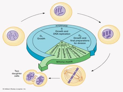
Interphase: The longest stage, where the cell grows, carries out normal functions, and prepares for division. Includes G1, S, and G2 phases.
Mitosis: Division of the nucleus to produce two identical daughter cells.
Cytokinesis: Division of the cytoplasm, resulting in two separate cells.
Meiosis: Specialized division producing gametes (sperm and eggs) with half the chromosome number.
Interphase
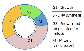
Interphase is the preparatory phase, taking up about 90% of the cell’s life cycle. It is divided into three stages:
G1 Phase (Gap 1): Cell grows and produces proteins, preparing for DNA replication. Normal cellular activities occur.
S Phase (Synthesis): DNA is replicated, forming sister chromatids in preparation for cell division.
G2 Phase (Gap 2): Cell continues to grow, produces proteins required for mitosis, duplicates organelles, and checks for DNA errors.
Mitosis
Stages of Mitosis
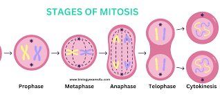
Mitosis is the process by which a single cell divides to produce two genetically identical daughter cells, maintaining chromosome number and genetic stability. The stages are:
Prophase: Chromosomes condense and become visible, each consisting of two sister chromatids joined at the centromere. The nuclear membrane breaks down and spindle fibers form.
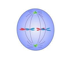
Metaphase: Chromosomes align along the cell’s equator, and spindle fibers attach to the centromeres, ensuring equal chromosome distribution.
Anaphase: Sister chromatids separate at the centromere and are pulled to opposite poles, ensuring genetic consistency in daughter cells.
Telophase: New nuclear membranes form around each set of chromosomes, which begin to uncoil. The cell prepares to split.
Cytokinesis: The cytoplasm divides, resulting in two identical daughter cells.

Meiosis
Overview and Significance
Meiosis is a specialized form of cell division that occurs in reproductive organs (testes and ovaries) to produce gametes (sperm and eggs). It involves two consecutive divisions, resulting in four genetically unique haploid cells, each with half the chromosome number of the parent cell.
Meiosis I: Homologous chromosomes are separated, reducing chromosome number by half. Crossing over occurs during Prophase I, exchanging genetic material between homologous chromosomes.
Meiosis II: Sister chromatids are separated, forming four unique haploid cells.
Meiosis increases genetic variation through crossing over and independent assortment, which is essential for evolution and species survival.
Comparison of Mitosis and Meiosis
Feature | Mitosis | Meiosis |
|---|---|---|
Location | Somatic (body) cells | Reproductive organs (testes, ovaries) |
Number of Divisions | One | Two (Meiosis I & II) |
Process Overview | DNA replicates once, cell divides once | DNA replicates once, cell divides twice |
Chromosome Number | Diploid → Diploid | Diploid → Haploid |
Genetic Variation | None (identical cells) | High (crossing over, independent assortment) |
Function | Growth, repair, replacement | Sexual reproduction (gamete formation) |
Type of Cells Produced | Somatic cells | Gametes (sperm, eggs) |
Product Outcome | Two identical diploid cells | Four unique haploid cells |
Significance | Genetic stability | Genetic diversity |
Summary
The cell cycle controls how new cells form, with interphase for growth and preparation, mitosis for identical cells, and meiosis for genetic diversity in reproduction.
Errors in the cell cycle can lead to diseases such as cancer, where cells divide uncontrollably due to failed regulation mechanisms.
