 Back
BackLESSON 1: Cells and Cell Membrane: Structure and Function
Study Guide - Smart Notes
 Tailored notes based on your materials, expanded with key definitions, examples, and context.
Cells and Cell Membrane: Structure and Function
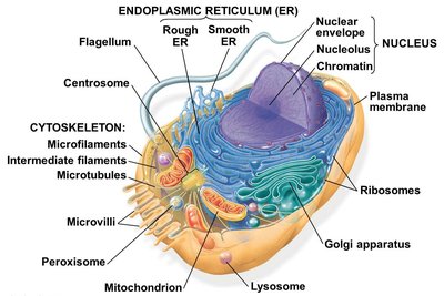
Overview of Animal Cell Structure
The animal cell is a complex unit composed of various organelles, each with specialized functions essential for cellular life. Understanding the structure and function of these organelles is foundational to cell biology.
Nucleus: Contains genetic material (DNA) and controls cellular activities.
Mitochondria: Site of cellular respiration and energy (ATP) production.
Endoplasmic Reticulum (ER): Rough ER is involved in protein synthesis; Smooth ER in lipid synthesis and detoxification.
Golgi Apparatus: Modifies, sorts, and packages proteins and lipids for secretion or delivery to other organelles.
Lysosomes: Contain digestive enzymes for breaking down waste.
Ribosomes: Sites of protein synthesis; not membrane-bound.
Cytoskeleton: Provides structural support and facilitates cell movement.
Plasma Membrane: Encloses the cell, regulating entry and exit of substances.
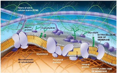
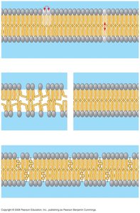
Cell Membrane Structure: The Fluid Mosaic Model
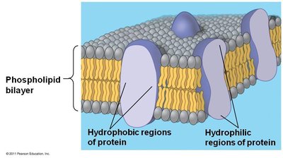
The plasma membrane is a selectively permeable barrier composed primarily of a phospholipid bilayer with embedded proteins, cholesterol, and carbohydrates. This structure is described by the fluid mosaic model, which highlights the dynamic and heterogeneous nature of the membrane.
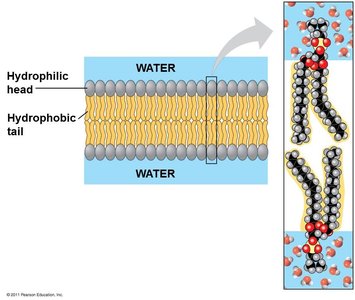
Phospholipid Bilayer: Consists of hydrophilic (water-attracting) heads and hydrophobic (water-repelling) tails, forming a double layer.
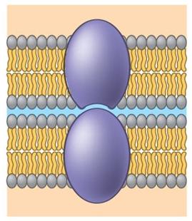
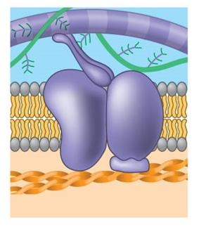
Membrane Proteins: Integral and peripheral proteins serve various functions, including transport, signaling, and structural support.
Cholesterol: Modulates membrane fluidity and stability.
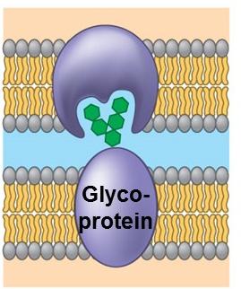
Carbohydrates: Attached to proteins (glycoproteins) or lipids (glycolipids) for cell recognition and signaling.
Membrane Fluidity
Membrane fluidity is crucial for proper membrane function, affecting the movement of proteins and lipids within the bilayer and the ability of the cell to adapt to changing conditions.
Lateral Movement: Phospholipids and some proteins can move laterally within the layer, contributing to fluidity.
Factors Affecting Fluidity:
Unsaturated hydrocarbon tails increase fluidity due to kinks that prevent tight packing.
Saturated hydrocarbon tails decrease fluidity by allowing tight packing.
Cholesterol acts as a fluidity buffer, restraining movement at high temperatures and preventing solidification at low temperatures.
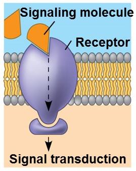
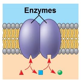
Functions of Membrane Proteins
Membrane proteins are essential for a variety of cellular processes. They are classified based on their roles:
Transport Proteins: Facilitate the movement of substances across the membrane, either passively or actively.
Enzymatic Activity: Catalyze specific reactions at the membrane surface.
Signal Transduction: Transmit signals from the external environment to the cell's interior.
Cell-Cell Recognition: Allow cells to identify each other, important in immune response.
Intercellular Joining: Connect adjacent cells for tissue formation.
Attachment to Cytoskeleton and Extracellular Matrix: Maintain cell shape and stabilize membrane proteins.

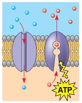
Transport Across the Membrane
The plasma membrane regulates the movement of substances into and out of the cell through various mechanisms:
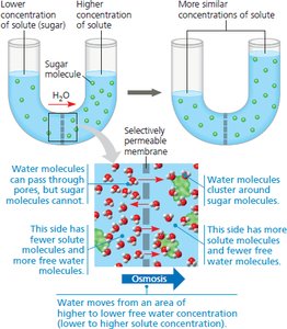
Passive Transport: Movement of molecules down their concentration gradient without energy input. Includes simple diffusion, facilitated diffusion (via channel or carrier proteins), and osmosis (water movement).
Active Transport: Movement of molecules against their concentration gradient, requiring energy (usually ATP). Example: sodium-potassium pump.
Bulk Transport: Large molecules (proteins, polysaccharides) are transported via vesicles in processes called endocytosis (into the cell) and exocytosis (out of the cell).
Summary Table: Types of Membrane Transport
Type | Energy Required? | Direction | Example |
|---|---|---|---|
Simple Diffusion | No | Down gradient | Oxygen, CO2 |
Facilitated Diffusion | No | Down gradient | Glucose, ions via channels |
Osmosis | No | Down water potential gradient | Water |
Active Transport | Yes (ATP) | Against gradient | Na+/K+ pump |
Bulk Transport | Yes (ATP) | In or out | Endocytosis, exocytosis |
Key Terms and Definitions
Phospholipid: A lipid molecule with a hydrophilic head and two hydrophobic tails, forming the basic structure of cell membranes.
Integral Protein: A protein embedded within the lipid bilayer, often spanning the membrane.
Peripheral Protein: A protein attached to the membrane surface.
Glycoprotein: A protein with carbohydrate chains attached, involved in cell recognition.
Osmosis: The diffusion of water across a selectively permeable membrane.
Endocytosis: The process of taking in large molecules by engulfing them in a vesicle.
Exocytosis: The process of expelling large molecules by fusing a vesicle with the plasma membrane.
Additional info:
Membrane fluidity is also influenced by temperature; lower temperatures decrease fluidity, while higher temperatures increase it.
Cells can adjust the composition of their membranes (e.g., increasing unsaturated fatty acids) to maintain optimal fluidity under different environmental conditions.
