 Back
BackCellular Reproduction: Mitosis, Meiosis, and Genetic Variation
Study Guide - Smart Notes
 Tailored notes based on your materials, expanded with key definitions, examples, and context.
Unit 5: Cellular Reproduction
Chapter 1: Cell Division
Cell division is a fundamental process by which cells reproduce, enabling growth, maintenance, and reproduction in living organisms. It can occur through asexual or sexual means, each with distinct biological implications.
Cell Division: The splitting of one parent cell into two genetically identical daughter cells. This process is essential for organismal growth, tissue repair, and reproduction in unicellular and multicellular organisms.
Asexual Reproduction: Involves cell division that results in the reproduction of a whole organism, producing clones—genetically identical individuals. Common in single-celled organisms and some multicellular organisms.
Sexual Reproduction: Requires the fusion of gametes (egg and sperm cells), resulting in offspring that are genetically distinct from their parents and each other. Gamete production occurs only in reproductive organs via a specialized cell division process.
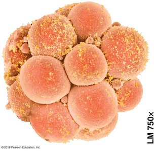
Cell Division in Multicellular Organisms: Sexual reproduction produces a zygote (fertilized egg), which undergoes repeated cell divisions to develop into an embryo and eventually a mature organism. Cell division also supports tissue renewal and repair.
Cell Division in Prokaryotes
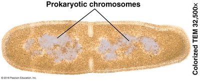
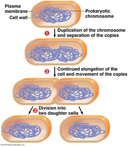
Prokaryotes, such as bacteria, reproduce by binary fission—a simpler process than eukaryotic cell division.
Binary Fission: "Dividing in half"—the prokaryotic chromosome (a single circular DNA molecule) is duplicated, and the cell splits into two identical daughter cells.
Eukaryotic Cell Division
Mitosis: Responsible for growth, maintenance, and asexual reproduction in multicellular organisms.
Meiosis: Produces gametes (egg and sperm) in reproductive organs for sexual reproduction.
Chapter 2: Mitosis
Mitosis is the process by which a eukaryotic cell divides its nucleus and genetic material, ensuring each daughter cell receives an identical set of chromosomes.
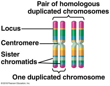
Eukaryotic Chromosomes
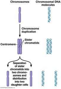
Chromosome: A single DNA molecule containing hundreds or thousands of genes. The number of chromosomes varies by species (e.g., humans have 46).
Chromatin: The complex of DNA and proteins that maintains chromosome structure and regulates gene expression. Chromatin condenses into visible chromosomes during cell division.
DNA Replication and Sister Chromatids
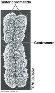
Before division, chromosomes are replicated, forming two sister chromatids joined at a region called the centromere.
During division, sister chromatids separate, ensuring each daughter cell receives an identical chromosome set.
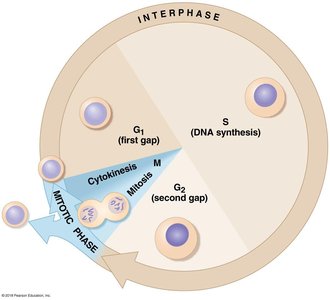
Cell Cycle
The cell cycle is an ordered sequence of events from cell formation to division. It consists of two main stages:
Interphase: The cell grows, performs normal functions, and replicates its DNA (G1, S, G2 phases).
Mitotic Phase (M Phase): Includes mitosis (nuclear division) and cytokinesis (cytoplasmic division).
Stages of Mitosis
Mitosis is divided into several stages, each with distinct events:
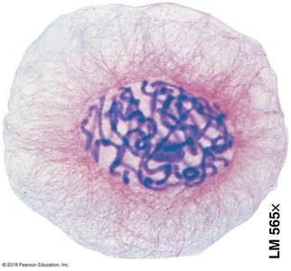
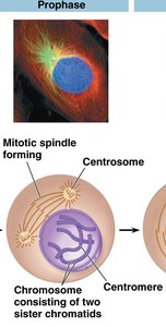
Prophase: Chromatin condenses into chromosomes; mitotic spindle forms from centrosomes.
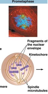
Prometaphase: Nuclear envelope fragments; spindle microtubules attach to kinetochores on chromosomes.
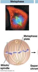
Metaphase: Chromosomes align at the metaphase plate; spindle is fully formed.
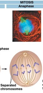
Anaphase: Sister chromatids separate and move toward opposite poles; cell elongates.
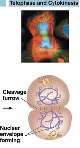
Telophase: Nuclear envelopes reform; chromosomes decondense; spindle disappears.

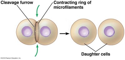
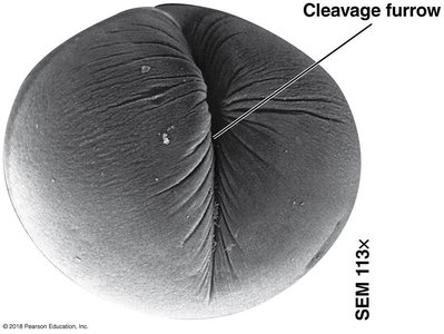
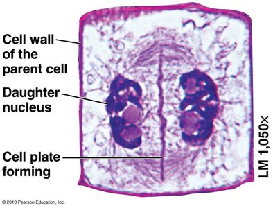
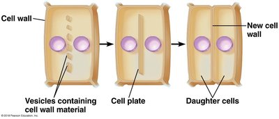
Cytokinesis: Division of the cytoplasm, resulting in two separate daughter cells.
Cytokinesis in Animal and Plant Cells
Animal Cells: A cleavage furrow forms, and a contractile ring of actin and myosin pinches the cell in two.
Plant Cells: Vesicles deliver cell wall materials to the center, forming a cell plate that develops into a new cell wall.
Regulation of Cell Division and Cancer
Anchorage Dependence: Most animal cells must be attached to a solid surface to divide.
Density-Dependent Inhibition: Cells stop dividing when crowded.
Growth Factors: Chemical signals required for cell division.
Cancer: Results from loss of cell cycle regulation, leading to uncontrolled cell growth and tumor formation. Tumors can be benign (localized) or malignant (invasive and metastatic).
Chapter 3: Meiosis
Meiosis is a specialized form of cell division that reduces the chromosome number by half, producing haploid gametes for sexual reproduction.
Types of Cells and Chromosomes
Somatic Cells: Typical body cells, diploid (2n), containing two sets of homologous chromosomes.
Gametes: Egg and sperm cells, haploid (n), containing one set of chromosomes.
Homologous Chromosomes: Chromosome pairs with the same genes but possibly different alleles.
Sex Chromosomes: Determine biological sex (X and Y in humans); autosomes are all other chromosomes.
Haploid vs. Diploid
Diploid (2n): Two sets of chromosomes (e.g., human somatic cells: 2n = 46).
Haploid (n): One set of chromosomes (e.g., human gametes: n = 23).
Life Cycle and Meiosis
The life cycle of sexually reproducing organisms alternates between diploid and haploid stages. Meiosis ensures the chromosome number is maintained across generations.
Stages of Meiosis
Interphase: Chromosomes duplicate, forming sister chromatids.
Meiosis I: Homologous chromosomes separate into two haploid cells (each chromosome still has two chromatids).
Meiosis II: Sister chromatids separate, resulting in four haploid daughter cells.
Chapter 4: Genetic Variation
Genetic variation arises from the processes of meiosis and fertilization, ensuring diversity in sexually reproducing populations.
Sources of Genetic Variation
Independent Assortment: The random orientation of homologous chromosome pairs during metaphase I leads to different combinations in gametes.
Random Fertilization: Any sperm can fertilize any egg, multiplying the possible genetic combinations.
Crossing Over: Exchange of genetic material between non-sister chromatids during prophase I creates recombinant chromosomes.
Chromosomal Abnormalities
Nondisjunction: Failure of chromosome pairs or sister chromatids to separate properly during meiosis, leading to gametes with abnormal chromosome numbers.
Karyotype: An ordered display of an individual's chromosomes, used to detect chromosomal abnormalities.
Down Syndrome: Caused by trisomy 21 (three copies of chromosome 21).
Sex Chromosome Abnormalities: Variations in X and Y chromosome number can affect development but are often less severe than autosomal abnormalities.
Alterations in Chromosome Structure
Deletion: Loss of a chromosome segment.
Duplication: Repetition of a chromosome segment.
Inversion: Reversal of a chromosome segment within the same chromosome.
Translocation: Segment moves to a non-homologous chromosome; can be reciprocal.
Clinical Relevance: Chromosomal alterations can cause birth defects and diseases such as cri-du-chat syndrome and chronic myelogenous leukemia (CML). Some changes have contributed to evolutionary processes.
Summary Table: Types of Chromosomal Alterations
Alteration | Description | Potential Effects |
|---|---|---|
Deletion | Loss of a chromosome segment | Often severe; e.g., cri-du-chat syndrome |
Duplication | Repetition of a segment | May cause developmental issues |
Inversion | Segment reversed within chromosome | Usually less harmful |
Translocation | Segment moves to another chromosome | May cause cancer or genetic disorders |
