 Back
BackChromosomes and Karyotypes: Structure, Organization, and Classification
Study Guide - Smart Notes
 Tailored notes based on your materials, expanded with key definitions, examples, and context.
Chromosomes: Structure and Organization
Genetic Material and the Genome
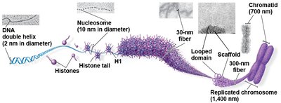
The genome of a cell comprises all the DNA present within it, which may be organized as a single DNA molecule (as in many prokaryotes) or as multiple DNA molecules (as in eukaryotes). The DNA is packaged with proteins to form structures called chromosomes.
Chromosome: A single, long DNA molecule associated with proteins, especially histones, that help in packaging and organization.
Chromatin: The complex of DNA and proteins that forms chromosomes within the nucleus of eukaryotic cells.
Nucleosome: The basic unit of DNA packaging, consisting of a segment of DNA wound around a core of histone proteins.
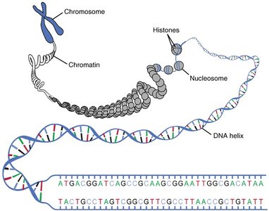
Example: Human cells contain approximately 2 meters of DNA per nucleus, compacted into chromosomes by histone proteins.
Bacterial and Organelle Genomes
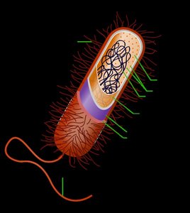
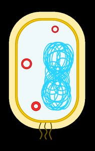
Prokaryotic (bacterial) genomes are typically composed of circular DNA molecules, which are supercoiled and localized in a region called the nucleoid. Eukaryotic organelles such as mitochondria and chloroplasts also contain small, circular DNA molecules, distinct from the nuclear genome.
Bacterial chromosome: Usually a single, circular DNA molecule with associated proteins.
Mitochondrial and chloroplast DNA: Small, circular DNA molecules found in these organelles, inherited maternally in most organisms.
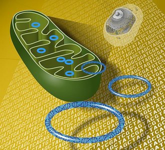
Example: Human mitochondria contain 37 genes essential for oxidative phosphorylation.
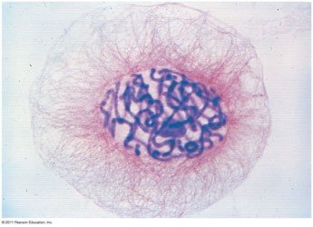
Eukaryotic Chromosomes
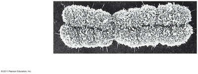
Eukaryotic cells typically possess multiple, linear chromosomes. During cell division, DNA is replicated and chromosomes condense, becoming visible under a microscope. Chromosomes are classified based on size, centromere position, and banding patterns after staining.
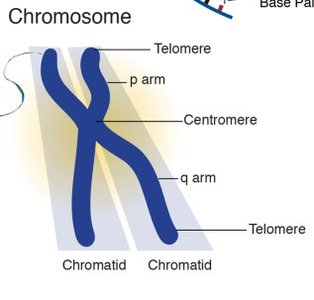
Centromere: The constricted region of a chromosome where sister chromatids are most closely attached.
Telomere: The repetitive DNA sequences at the ends of linear chromosomes, protecting them from degradation.
Sister chromatids: Identical copies of a chromosome, joined at the centromere, produced during DNA replication.
Classification of Chromosomes by Centromere Position
Chromosomes are classified based on the position of the centromere:
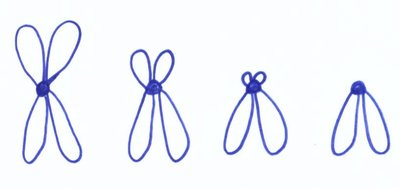
Metacentric: Centromere is in the middle, arms are of equal length.
Submetacentric: Centromere is slightly off-center, creating arms of unequal length.
Acrocentric: Centromere is near one end, producing a very short (p) arm and a long (q) arm.
Telocentric: Centromere is at the very end (not found in humans).
Karyotypes and Chromosome Grouping
Human Karyotype and Chromosome Groups
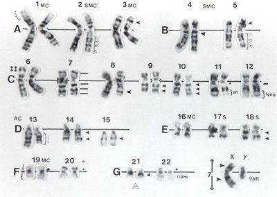
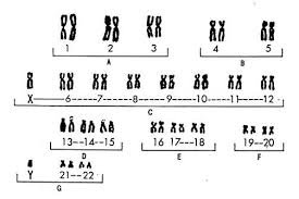
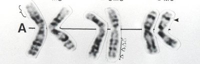
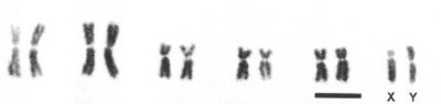
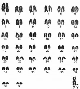
A karyotype is an ordered display of the chromosomes from a cell, arranged in pairs by size, centromere position, and banding pattern. Human somatic cells contain 23 pairs of chromosomes (46 total), grouped into seven classes (A–G) based on size and centromere position.
Group | Chromosomes | Size | Centromere Position |
|---|---|---|---|
A | 1–3 | Large | Submetacentric, Metacentric |
B | 4, 5 | Large | Submetacentric |
C | 6–12, X | Medium | Submetacentric |
D | 13–15 | Medium | Acrocentric |
E | 16–18 | Short | Submetacentric, Metacentric |
F | 19, 20 | Short | Metacentric |
G | 21, 22, Y | Very short | Acrocentric |
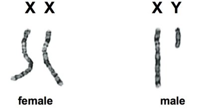
Homologous Chromosomes and Sex Chromosomes
Each chromosome pair consists of homologous chromosomes (homologs), which are the same length and shape and carry genes controlling the same inherited traits. Humans have 22 pairs of autosomes and one pair of sex chromosomes (XX for females, XY for males).
Autosomes: Chromosomes not involved in sex determination (pairs 1–22 in humans).
Sex chromosomes: X and Y chromosomes, determining biological sex.
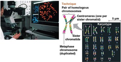
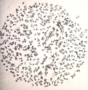
Karyotype Preparation and Staining
Karyotype analysis involves harvesting somatic cells, stimulating them to divide, arresting them in metaphase, staining the chromosomes, and photographing them under a microscope. Giemsa stain is commonly used, producing characteristic G bands that help identify individual chromosomes.
G bands: Dark and light bands on chromosomes after Giemsa staining, reflecting regions of differing DNA composition and structure.
Example: Karyotyping is used in clinical genetics to diagnose chromosomal abnormalities such as Down syndrome (trisomy 21).
Chromosome Numbers in Different Organisms
Species Variation in Chromosome Number
Each eukaryotic species has a characteristic number of chromosomes in its somatic cells. Most DNA is nuclear, but small amounts are found in mitochondria and chloroplasts. Chromosome numbers vary widely among species.
Organism | Chromosome Number (2n) |
|---|---|
Human | 46 |
Chicken | 78 |
Cat | 38 |
Sweet orange | 18 |
Ophioglossum reticulatum (fern) | 1262 |
Example: The fern Ophioglossum reticulatum has the highest known chromosome number among living organisms (2n = 1262).
Additional info: Chromosome structure and number are fundamental to understanding genetics, heredity, and cell division processes such as mitosis and meiosis.
