 Back
Ch: 34 Circulatory System: Structure, Function, and Physiology
Study Guide - Smart Notes
 Tailored notes based on your materials, expanded with key definitions, examples, and context.
Circulatory System Overview
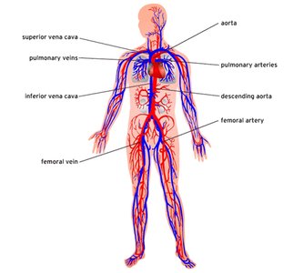
Functions of the Circulatory System
The circulatory system is essential for transporting nutrients, gases, hormones, and waste products throughout the body. It also plays a critical role in regulating body temperature, pH, and fluid balance, as well as protecting the body through immune responses and clotting mechanisms.
Transportation: Delivers oxygen and nutrients to tissues, removes carbon dioxide and metabolic wastes.
Regulation: Maintains homeostasis of temperature, pH, and fluid volume.
Protection: Involves immune cells and clotting factors to defend against pathogens and prevent blood loss.
Types of Circulatory Systems
Open vs. Closed Circulatory Systems
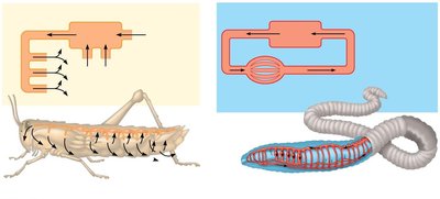
Animals have evolved two main types of circulatory systems: open and closed. The structure and function of these systems reflect the metabolic needs and complexity of the organism.
Open Circulatory System: Hemolymph bathes organs directly in an open cavity; found in arthropods and most mollusks.
Closed Circulatory System: Blood is confined to vessels and is distinct from interstitial fluid; found in annelids, cephalopods, and all vertebrates.
Single vs. Double Circulation
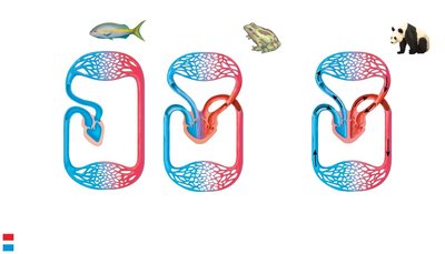
Vertebrates exhibit either single or double circulation patterns, which influence the efficiency of oxygen delivery and separation of oxygenated and deoxygenated blood.
Single Circulation: Blood passes through the heart once per circuit (e.g., fish).
Double Circulation: Blood passes through the heart twice per circuit (e.g., amphibians, reptiles, mammals).
Examples of Circulatory Systems in Vertebrates
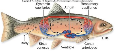
Fish: Two-chambered heart (one atrium, one ventricle); blood flows from heart to gills to body and back to heart.
Amphibians: Three-chambered heart (two atria, one ventricle); double circulation with some mixing of blood.
Mammals, Birds, Crocodilians: Four-chambered heart (two atria, two ventricles); complete separation of oxygenated and deoxygenated blood.
Vertebrate Heart: Structure and Physiology
Heart Chambers and Blood Flow
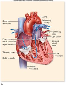
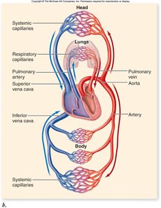
The vertebrate heart is a muscular organ that pumps blood through the circulatory system. Mammals and birds have a four-chambered heart, which ensures efficient separation of oxygen-rich and oxygen-poor blood.
Right Atrium: Receives deoxygenated blood from the body.
Right Ventricle: Pumps deoxygenated blood to the lungs.
Left Atrium: Receives oxygenated blood from the lungs.
Left Ventricle: Pumps oxygenated blood to the body.
The Cardiac Cycle
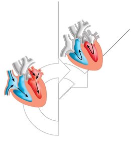
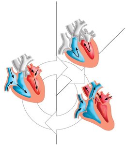
The cardiac cycle describes the sequence of events during one heartbeat, including periods of contraction (systole) and relaxation (diastole).
Diastole: Ventricles relax and fill with blood.
Systole: Ventricles contract and pump blood out of the heart.
Heart Sounds: "Lub" (closure of AV valves), "Dub" (closure of semilunar valves).
Electrophysiology of the Heart
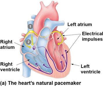
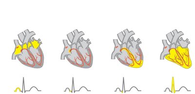
The heart contains autorhythmic fibers that generate and conduct electrical impulses, coordinating the heartbeat. The sinoatrial (SA) node acts as the natural pacemaker.
SA Node: Initiates electrical impulses, causing atria to contract.
AV Node: Delays impulse, allowing atria to empty before ventricles contract.
Bundle Branches and Purkinje Fibers: Distribute impulse through ventricles, causing contraction.
Autonomic Nervous System: Modulates heart rate via sympathetic (increases rate) and parasympathetic (decreases rate) input.
Pacemakers and Heart Rhythm
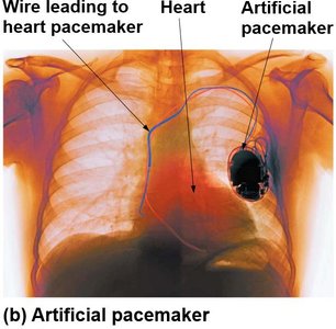
Disruptions in the heart's electrical system can cause arrhythmias. Artificial pacemakers can be implanted to restore normal rhythm.
Blood Vessels: Structure and Function
Types of Blood Vessels
Blood vessels form a closed network for blood flow. They vary in structure and function:
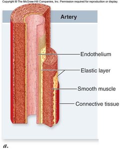
Arteries: Thick-walled, muscular vessels that carry blood away from the heart.
Arterioles: Small branches of arteries that regulate blood flow to capillaries.
Capillaries: Thin-walled vessels where gas and nutrient exchange occurs.
Venules: Small vessels that collect blood from capillaries.
Veins: Thin-walled vessels that return blood to the heart; contain valves to prevent backflow.
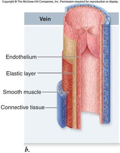
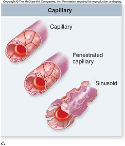
Blood Flow and Pressure
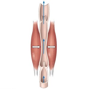
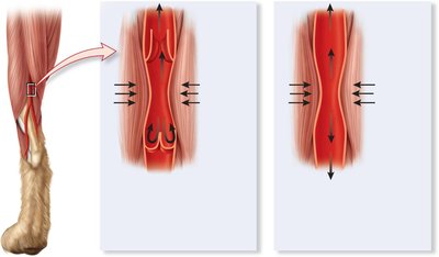
Blood pressure is highest in arteries and decreases through arterioles, capillaries, venules, and veins. Veins rely on skeletal muscle contractions and valves to return blood to the heart.
Blood Components
Plasma and Formed Elements
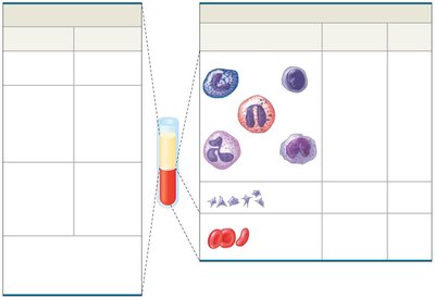
Blood is a connective tissue composed of a liquid matrix (plasma) and cellular elements (formed elements).
Plasma: Water, ions, proteins (albumin, fibrinogen, immunoglobulins), nutrients, hormones, and waste products.
Formed Elements: Erythrocytes (red blood cells), leukocytes (white blood cells), and platelets (cell fragments).
Blood Clotting
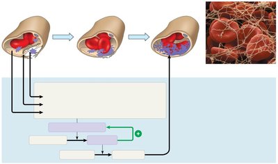
Platelets and plasma proteins are essential for blood clotting. The clotting cascade involves a series of enzymatic reactions that convert fibrinogen to fibrin, forming a stable clot.
Platelets: Cell fragments that initiate clot formation by adhering to damaged vessels.
Clotting Factors: Plasma proteins (including fibrinogen) and ions (e.g., calcium) that participate in the cascade.
Fibrin: Insoluble protein that forms the meshwork of a blood clot.
Hormonal Regulation of Blood Volume and Pressure
Blood volume and pressure are regulated by hormones such as antidiuretic hormone (ADH), aldosterone, and atrial natriuretic hormone, which influence kidney function and water retention.
ADH: Promotes water reabsorption in kidneys, increasing blood volume.
Aldosterone: Increases sodium and water retention, raising blood pressure.
Atrial Natriuretic Hormone: Reduces blood volume and pressure by promoting sodium excretion.
