 Back
Diabetes Unit: Digestion, Transport, Endocrine System, and Cellular Respiration Study Guide
Study Guide - Smart Notes
 Tailored notes based on your materials, expanded with key definitions, examples, and context.
Digestion and Transport
Digestive Organs and Their Functions
The digestive system is composed of specialized organs that break down food into absorbable nutrients. Each organ has a unique function in the process of digestion and nutrient absorption.
Mouth: Mechanical breakdown and enzymatic digestion (amylase for carbohydrates).
Stomach: Protein digestion via pepsin; acidic environment denatures proteins.
Small Intestine: Major site for digestion and absorption; enzymes from pancreas and bile from liver aid in breakdown.
Large Intestine: Water absorption and formation of feces.
Food Passage: Food passes through the mouth, esophagus, stomach, small intestine, large intestine, and exits via the rectum.
Macromolecules in Foods
Foods contain different macromolecules, each with distinct properties and nutritional roles.
Carbohydrates: Found in grains, fruits, vegetables; provide energy.
Proteins: Found in meat, dairy, legumes; essential for growth and repair.
Fats/Lipids: Found in oils, butter, nuts; energy storage and cell membrane structure.
Nucleic Acids: Present in all cells; not a major dietary component.
Structure and Function in Nutrient Absorption
The structure of digestive organs and cell membranes determines how nutrients are absorbed. The polarity of molecules affects their transport across membranes.
Polar molecules (hydrophilic) require transport proteins to cross cell membranes.
Non-polar molecules (hydrophobic) can diffuse directly through lipid bilayers.
Enzymes and Their Function
Enzymes are biological catalysts that speed up chemical reactions in digestion. They are specific to substrates and lower activation energy.
Example: Amylase breaks down starch into glucose.
Transport Mechanisms
Transport across cell membranes can be active or passive.
Passive Transport: Diffusion and facilitated diffusion; no energy required.
Active Transport: Requires energy (ATP); moves molecules against concentration gradient.
Transmembrane Proteins: Channels and carriers for passive transport; pumps for active transport.
Polarity and Membrane Transport
The polarity of monomers determines their solubility and transport.
Polar bonds: Often involve oxygen or nitrogen (e.g., O-H, N-H).
Non-polar bonds: C-H bonds are most common.
Structures aiding transport: Carrier proteins, channel proteins, and pumps.
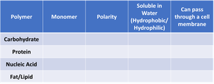
Polymer | Monomer | Polarity | Soluble in Water (Hydrophobic/Hydrophilic) | Can pass through a cell membrane |
|---|---|---|---|---|
Carbohydrate | Monosaccharide | Polar | Hydrophilic | No (requires transport proteins) |
Protein | Amino Acid | Polar/Non-polar (varies) | Mostly hydrophilic | No (requires transport proteins) |
Nucleic Acid | Nucleotide | Polar | Hydrophilic | No (requires transport proteins) |
Fat/Lipid | Fatty Acid/Glycerol | Non-polar | Hydrophobic | Yes (can diffuse through membrane) |
Cell Membranes
Components and Properties
Cell membranes are composed of a phospholipid bilayer with embedded proteins, cholesterol, and carbohydrates. Their structure determines permeability and transport mechanisms.
Phospholipids: Amphipathic molecules with hydrophilic heads and hydrophobic tails.
Proteins: Channels, carriers, receptors.
Cholesterol: Modulates fluidity.
Diffusion vs. Facilitated Diffusion: Diffusion is passive movement of molecules; facilitated diffusion uses proteins to help polar molecules cross.
Endocrine System
Hormones and Their Transport
Hormones are signaling molecules that regulate physiological processes. Their polarity affects their transport, receptor interaction, and movement in the blood.
Types: Amine, peptide/protein, lipid/steroid.
Polarity: Peptide/protein hormones are polar; steroid hormones are non-polar.
Transport: Polar hormones require blood transporters; non-polar hormones can diffuse through membranes.
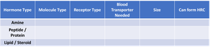
Hormone Type | Molecule Type | Receptor Type | Blood Transporter Needed | Size | Can form HRC |
|---|---|---|---|---|---|
Amine | Small, polar | Membrane receptor | No | Small | No |
Peptide/Protein | Large, polar | Membrane receptor | Yes | Large | No |
Lipid/Steroid | Non-polar | Intracellular receptor | Yes | Small/medium | Yes |
Transport vs. Transduction
Transport refers to the movement of hormones into cells, while transduction is the process of signal relay via receptors. Steroid hormones are transported into cells; peptide hormones initiate signal transduction at the membrane.
Transported: Steroid hormones.
Signal Transduction: Peptide/protein hormones.
Endocrine vs. Nervous System
The endocrine system uses hormones for slow, sustained signaling; the nervous system uses electrical impulses for rapid, targeted responses.
Pros (Endocrine): Long-lasting, widespread effects.
Cons (Endocrine): Slow response.
Pros (Nervous): Fast, precise.
Cons (Nervous): Short duration, limited scope.
Dose Response
Dose response describes how the magnitude of hormone response changes with hormone concentration.
Example: Higher insulin levels increase glucose uptake until saturation.
Diabetes and Glucose Regulation
Feedback Mechanisms
Homeostasis is maintained by negative and positive feedback loops. Negative feedback returns a system to its set point; positive feedback amplifies changes.
Negative Feedback: Blood glucose regulation.
Positive Feedback: Blood clotting.
Blood Glucose Regulation
Blood glucose is regulated by hormones released from the pancreas.
High Blood Sugar: Insulin released; promotes glucose uptake and storage.
Low Blood Sugar: Glucagon released; promotes glucose release from liver.
Hyperglycemic: High blood glucose; Hypoglycemic: Low blood glucose.
Roles in Homeostasis
Receptors: Detect blood glucose levels.
Control Centers: Pancreas.
Effectors: Liver, muscle, fat cells.
Liver's Role
The liver stores glucose as glycogen and releases it when needed.
Hyperglycemic: Stores glucose.
Hypoglycemic: Releases glucose.
Insulin and GLUT-4
Insulin allows glucose to enter cells by promoting the insertion of GLUT-4 transporters into the cell membrane. GLUT-4 is regulated and only active when insulin is present.
Types of Diabetes
Type 1: Autoimmune destruction of insulin-producing cells.
Type 2: Insulin resistance; cells do not respond to insulin.
Diagnosis: Blood glucose tests, HbA1c.
Treatment: Type 1: Insulin therapy; Type 2: Lifestyle changes, medications.
Public Health: Focuses on prevention and behavior change; doctors focus on individual treatment.
Hormone Regulation
Dose Response and Hormone Axes
Hormone axes involve stimulating and suppressing hormones. Upregulation increases receptor number, amplifying response; downregulation decreases receptor number, dampening response.
Competitive Inhibitors: Block hormone binding at receptor.
Allosteric Inhibitors: Change receptor shape, reducing hormone effect.
Hormone Interactions: Synergistic, antagonistic, permissive.
Respiration
Mitochondria and Cellular Respiration
Mitochondria are organelles found in eukaryotes; they are the site of cellular respiration.
Stages: Glycolysis, Pyruvate Oxidation, Citric Acid Cycle, Oxidative Phosphorylation.
Take Home Message: Energy from glucose is converted to ATP.
Oxidation and Reduction
Cellular respiration involves oxidation (loss of electrons) and reduction (gain of electrons). These reactions are always paired.
Glucose: Oxidized to CO2.
Oxygen: Reduced to H2O.
Key Equation:
ATP Production
ATP is produced via substrate-level phosphorylation and oxidative phosphorylation. The electron transport chain (ETC) and ATP synthase are essential for ATP generation.
FAD/FADH2 and NAD+/NADH: Electron carriers in respiration.
ATP Yield: Glycolysis (2 ATP), Citric Acid Cycle (2 ATP), Oxidative Phosphorylation (~34 ATP).
Additional info: Some explanations and table entries were inferred for completeness and clarity based on standard biology curriculum.
