 Back
BackFlowering Plant Anatomy: Structure, Function, and Diversity of Plant Cells and Tissues
Study Guide - Smart Notes
 Tailored notes based on your materials, expanded with key definitions, examples, and context.
Flowering Plant Anatomy
Introduction to Plant Anatomy
Plant anatomy is the study of the internal structure of plants, focusing on the organization and function of cells and tissues. Understanding plant anatomy is essential for comprehending how plants grow, transport materials, and interact with their environment.
Meristematic Tissue
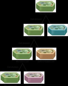
Meristem: Plant Stem Cells
Meristematic cells are undifferentiated cells found in specific regions of plants, responsible for growth and regeneration. These cells can divide continuously, producing new cells that either remain meristematic or differentiate into specialized cell types.
Meristematic cells: Undifferentiated, capable of division and differentiation.
Apical meristem: Located at root and shoot tips, responsible for primary growth (lengthening).
Lateral meristem: Responsible for secondary growth (thickening), includes vascular cambium and cork cambium.
Dedifferentiation: Specialized cells can revert to a meristematic state under certain conditions.
Indeterminate growth: Plants retain meristematic tissue throughout life, allowing continuous growth.
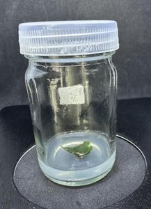
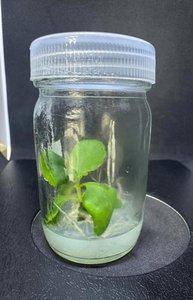
Plant Tissue Culture
Plant tissue culture exploits the ability of plant cells to dedifferentiate and regenerate into whole plants under suitable conditions. This technique is widely used in research and agriculture for cloning and genetic modification.
Dedifferentiation: Cells revert to meristematic state.
Regeneration: Meristematic cells can develop into a complete plant.
Applications: Cloning, genetic engineering, conservation.
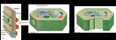
Plant Cell Wall
Primary and Secondary Cell Walls
All plant cells possess a primary cell wall, which is flexible and allows for growth. Some cells develop a secondary cell wall, which is rigid and provides structural support.
Primary cell wall: Thin, flexible, allows cell expansion.
Secondary cell wall: Thick, rigid, contains lignin, provides strength.
Lignin: Complex polymer that strengthens the secondary wall, stains red with safranin O.
Types of Plant Cells
Parenchyma Cells
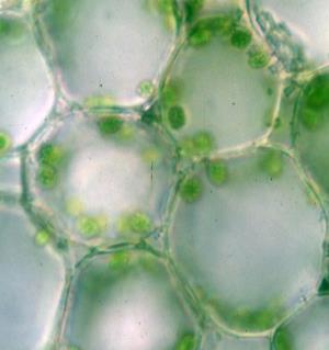
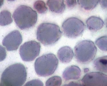
Parenchyma cells are the most common and versatile plant cells, involved in photosynthesis, storage, and metabolic functions. They have thin primary walls and are alive at maturity.
Shape: Usually spherical or polyhedral.
Functions: Photosynthesis (chlorenchyma), storage (aerenchyma, starch), lateral transport.
Specialized types: Chlorenchyma (with chloroplasts), aerenchyma (with air spaces).
Collenchyma Cells
Collenchyma cells provide structural support in young plant tissues. They have unevenly thickened primary walls and are alive at maturity, allowing flexibility and strength.
Wall thickness: Thick primary walls, especially at corners.
Function: Support in growing regions (stems, petioles).
Appearance: White and shiny under microscope.
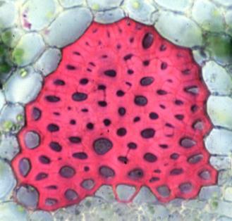
Sclerenchyma Cells
Sclerenchyma cells are dead at maturity and have thick secondary walls containing lignin. They provide structural support and are classified into fibers and sclereids.
Fibers: Elongated cells, found in vascular bundles.
Sclereids: Irregularly shaped, found in hard plant parts (e.g., seed coats, gritty texture in fruits).
Function: Mechanical support.
Water Conducting Cells: Tracheids and Vessel Members
Tracheids and vessel members are specialized for water transport in the xylem. Both are dead at maturity and have secondary walls.
Tracheids: Narrow, tapered ends, found in all vascular plants.
Vessel members: Wide, blunt ends with perforation plates, found in angiosperms.
Function: Water transport and support.
Food Conducting Cells: Sieve Tube Members
Sieve tube members are elongated cells in the phloem, responsible for transporting food. They are alive at maturity but lack a nucleus, and are accompanied by companion cells.
Sieve tube members: Transport sugars and other organic molecules.
Companion cells: Support sieve tube members metabolically.
Sieve plates: Porous end walls facilitating transport.
Epidermal Cells and Guard Cells
Epidermal cells form the outermost layer of plant tissues, providing protection. Guard cells regulate the opening and closing of stomata, controlling gas exchange and water loss.
Epidermal cells: Outermost layer, thick cuticle.
Guard cells: Control stomatal aperture.
Subsidiary cells: Surround guard cells, may be absent in some plants.
Plant Tissue Organization
Primary Growth and Tissue Types
Primary growth increases the length of roots and shoots, accomplished by apical meristems. Plant tissues are organized into three main types: epidermal, vascular, and ground tissue.
Epidermal tissue: Protection and gas exchange.
Vascular tissue: Xylem (water transport), phloem (food transport).
Ground tissue: Photosynthesis, storage, support.
Simple tissue: Composed of one cell type.
Complex tissue: Composed of multiple cell types.
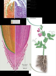
Root Anatomy
Roots exhibit three zones of growth: cell division, elongation, and maturation. Root hairs increase surface area for absorption. Monocot and dicot roots differ in stele organization.
Zone of cell division: Contains apical meristem.
Zone of elongation: Cells elongate, pushing root tip forward.
Zone of maturation: Cells differentiate, root hairs form.
Pericycle: Origin of lateral roots in dicots.
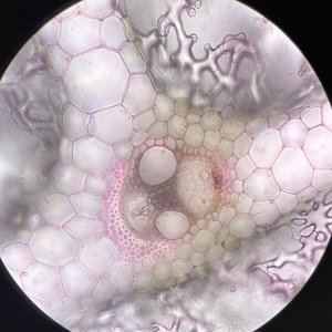
Stem Anatomy
Stems contain vascular bundles, ground tissue, and epidermis. Monocot stems have scattered vascular bundles, while dicot stems have bundles arranged in a ring.
Monocot stem: Scattered vascular bundles.
Dicot stem: Bundles in a ring, with vascular cambium for secondary growth.
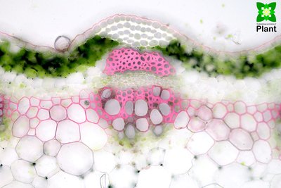
Leaf Anatomy
Leaves are organized into epidermis, mesophyll (palisade and spongy), and vascular bundles. Mesophyll consists of parenchyma cells specialized for photosynthesis.
Palisade mesophyll: Columnar parenchyma cells, high photosynthetic activity.
Spongy mesophyll: Loosely arranged parenchyma cells, gas exchange.
Vascular bundle: Contains xylem and phloem.
Secondary Growth
Vascular Cambium and Cork Cambium
Secondary growth increases the thickness of stems and roots, produced by lateral meristems. Vascular cambium generates secondary xylem (wood) and phloem, while cork cambium produces cork for protection.
Vascular cambium: Produces secondary xylem and phloem.
Cork cambium: Produces cork, part of bark.
Annual rings: Early wood (large cells, light color), late wood (small cells, dark color).
Lenticels: Openings in bark for gas exchange.
Heartwood: Non-functional secondary xylem.
Sapwood: Functional secondary xylem.
Summary Table: Types of Plant Cells
Cell Type | Wall Type | Alive at Maturity? | Main Function | Special Features |
|---|---|---|---|---|
Meristematic | Primary | Yes | Growth, division | Undifferentiated |
Parenchyma | Primary | Yes | Photosynthesis, storage, metabolism | Versatile, various shapes |
Collenchyma | Thickened primary | Yes | Support in young tissues | Flexible, uneven walls |
Sclerenchyma (Fiber/Sclereid) | Secondary | No | Support | Lignified, rigid |
Tracheid/Vessel | Secondary | No | Water transport | Dead, hollow |
Sieve Tube Member | Primary | Yes (no nucleus) | Food transport | Sieve plates, companion cells |
Epidermal/Guard | Primary | Yes | Protection, gas exchange | Cuticle, stomata |
Conclusion
Understanding the diversity of plant cells and tissues is fundamental to biology. Each cell type and tissue plays a specific role in plant growth, structure, and function, enabling plants to thrive in diverse environments.
