 Back
BackHistology: Structure and Function of Animal Tissues
Study Guide - Smart Notes
 Tailored notes based on your materials, expanded with key definitions, examples, and context.
Histology: The Study of Plant and Animal Tissues
Introduction to Histology
Histology is the branch of biology concerned with the microscopic study of tissue structure and function in plants and animals. Understanding tissue types is fundamental for recognizing how form relates to function in living organisms.
Tissue: A group of cells with similar structure and function.
Form follows function: The structure of a biological component is shaped by its functional requirements.
Hierarchy of Structural Organization in Animals
Animals are organized in a hierarchical manner, from cells to tissues, organs, and organ systems.
Cell: Basic unit of life.
Tissue: Groups of similar cells performing a specific function.
Organ: Structure composed of multiple tissue types working together.
Organ system: Group of organs performing major body functions.
Main Tissue Categories
Epithelial Tissue
Epithelial tissues cover body surfaces, line internal cavities, and form glands. Their structure is closely related to their protective, absorptive, and secretory functions.
Columnar epithelium: Tall, column-shaped cells found in the stomach and intestines; specialized for absorption and secretion.
Ciliated pseudostratified columnar epithelium: Appears layered but all cells touch the basement membrane; cilia help move mucus in the trachea and upper respiratory tract.
Simple cuboidal epithelium: Cube-shaped cells, often found in kidney tubules; involved in secretion and absorption.
Connective Tissue
Connective tissues provide structural support, store energy, and connect other tissues.
Red and white blood cells (erythrocytes and leukocytes): Blood is a fluid connective tissue; erythrocytes transport oxygen, leukocytes defend against pathogens.
Bone (compact): Dense, rigid tissue providing structural support and protection.
Adipose tissue: Stores fat for energy and insulation.
Hyaline cartilage: Smooth, glassy tissue found in joints, providing cushioning.
Loose (areolar) connective tissue: Flexible tissue that binds organs and supports blood vessels.
Aorta (elastic): Elastic connective tissue in large arteries allows for stretch and recoil.
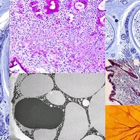
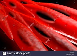
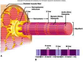
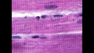
Muscle Tissue
Muscle tissue is specialized for contraction and movement. It is the most abundant tissue in animals and contains contractile proteins actin and myosin.
Skeletal muscle: Attaches to bones via tendons, responsible for voluntary movement, and has a striated appearance due to organized contractile units.
Cardiac muscle: Found only in vertebrate hearts, striated but involuntary, with branched fibers and intercalated discs for rapid signal transmission.
Smooth muscle: Lacks striations, spindle-shaped cells, contracts slowly, found in walls of digestive tract, arteries, and internal organs; responsible for involuntary movements like peristalsis.

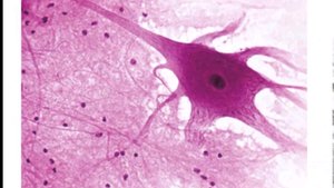
Nervous Tissue
Nervous tissue senses stimuli and rapidly transmits information throughout the body.
Neuron: Functional unit of nervous tissue, conducts electrical impulses.
Glia: Supporting cells that insulate, nourish, and regulate the environment for neurons.

Organs and Organ Systems
Organs
Organs are composed of multiple tissue types arranged in layers to perform specific functions. The integration of tissues allows organs to carry out complex tasks that individual tissues cannot accomplish alone. 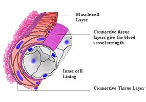
Organ Systems
Organ systems are groups of organs that work together to perform major physiological functions, such as digestion, circulation, and respiration.
Example: The circulatory system includes the heart (cardiac muscle), blood vessels (smooth muscle and connective tissue), and blood (connective tissue).
Summary Table: Main Tissue Types and Features
Tissue Type | Main Features | Location |
|---|---|---|
Epithelial | Protective, absorptive, secretory; tightly packed cells | Skin, lining of organs |
Connective | Support, storage, transport; diverse cell types in matrix | Bone, blood, cartilage, fat |
Muscle | Contractile, movement; elongated fibers | Skeletal muscles, heart, digestive tract |
Nervous | Signal transmission; neurons and glia | Brain, spinal cord, nerves |
Additional info:
Distinguishing between lymphocytes and neutrophils: Lymphocytes have a large, round nucleus and little cytoplasm; neutrophils have a multi-lobed nucleus and more cytoplasm.
Intercalated discs in cardiac muscle: Specialized junctions that facilitate rapid signal transmission for synchronized heart contraction.
Peristalsis: Wave-like contractions of smooth muscle in the digestive tract that move food along.
