 Back
BackHistology: Structure and Function of Animal Tissues
Study Guide - Smart Notes
 Tailored notes based on your materials, expanded with key definitions, examples, and context.
Histology: The Study of Animal Tissues
Introduction to Histology
Histology is the branch of biology that studies the microscopic structure of tissues in plants and animals. Understanding tissue structure is essential for recognizing how form relates to function in biological systems. - Tissue: A group of cells with a common structure and function. - Form follows function: The anatomical structure of a tissue or organ is directly related to its physiological role. 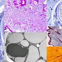
Hierarchy of Structural Organization in Animals
Biological structure is organized in a hierarchy, from cells to tissues, organs, organ systems, and the whole organism. - Cell: Basic unit of life. - Tissue: Group of similar cells performing a specific function. - Organ: Structure composed of multiple tissue types working together. - Organ system: Group of organs performing related functions. - Organism: Complete living entity. 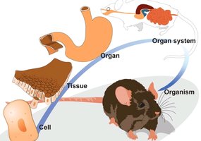
Epithelial Tissue
General Features of Epithelial Tissue
Epithelial tissues are sheets of closely packed cells that cover body surfaces and line internal organs. They form protective barriers and facilitate exchange with tissue fluid, blood, or air. - Apical surface: Faces outside of organ or inside of tube/passageway. - Basal lamina: Matrix separating epithelium from underlying tissue. 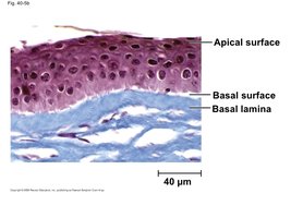
Classification of Epithelial Tissue
Epithelial tissues are classified by the number of cell layers and the shape of cells on the apical surface. - Simple: One layer of cells. - Stratified: Multiple layers. - Cell shapes: Squamous (flat), cuboidal (cube-shaped), columnar (tall). 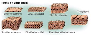
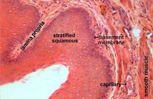
Squamous Epithelium
Squamous cells are thin and leaky, suitable for diffusion. - Simple squamous: Lines capillaries and alveoli for rapid exchange. - Stratified squamous: Protects surfaces subject to abrasion (e.g., mouth, esophagus). 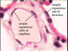
Cuboidal Epithelium
Cuboidal cells have large cytoplasmic volume for secretion and absorption. - Found in kidney tubules and glands. 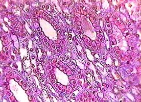
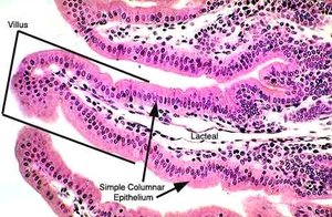
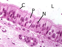
Columnar and Pseudostratified Epithelium
Columnar cells are specialized for secretion and absorption. - Simple columnar: Lines intestines for nutrient absorption and digestive juice secretion. - Pseudostratified columnar: Ciliated, forms mucous membrane in respiratory tract for trapping/removal of particles. 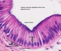
Connective Tissue
General Features of Connective Tissue
Connective tissue consists of cells scattered in an extracellular matrix, which may be liquid, jelly-like, or solid. The matrix is produced by the cells and contains fibers. 
Loose Connective Tissue
The most widespread connective tissue, with a loose weave of fibers. - Contains strong collagen and elastic fibers. - Binds epithelia to underlying tissue and holds organs in place. 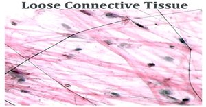
Fibrous Connective Tissue
Contains densely packed bundles of collagen fibers for maximum strength. - Forms tendons (muscle to bone) and ligaments (bone to joint). 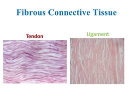
Adipose Tissue
Stores fat, pads and insulates the body, and stores energy. - Adipose cells contain fat droplets that swell or shrink. 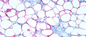
Cartilage
Cartilage is strong and flexible, with collagen fibers in a rubbery matrix. - Provides shock absorption and support (e.g., ends of bones, ears, nose). 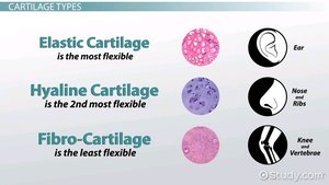
Bone
Bone has a matrix of collagen fibers embedded in hard minerals (Ca, Mg, PO4). - Strong but not brittle; contains living cells in circular layers for growth and repair. 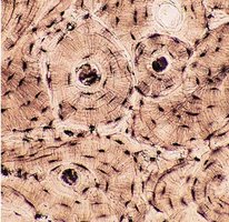
Blood
Blood is a connective tissue with a liquid matrix (plasma) and suspended formed elements (cells and platelets). - Main transport system in the body. 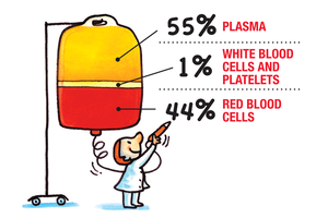
Blood Cells
- White blood cells (leukocytes): Defend against disease. - Red blood cells (erythrocytes): Carry O2, H+, CO2. - Platelets (thrombocytes): Aid in blood clotting. 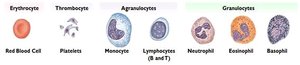
Summary Table: Major Animal Tissue Types
Tissue Type | Main Function | Key Features |
|---|---|---|
Epithelial | Protection, absorption, secretion | Sheets of cells, apical/basal surfaces |
Connective | Support, binding, transport | Cells in matrix, fibers (collagen, elastic) |
Muscle | Movement | Contractile cells |
Nervous | Communication | Neurons and glial cells |
Additional info:
- Muscle and nervous tissues are also covered in histology but not detailed in this excerpt. - The structure of each tissue type is closely related to its function, as illustrated by the examples above.
