 Back
BackInside the Cell: Structure, Function, and Dynamics of Cellular Components
Study Guide - Smart Notes
 Tailored notes based on your materials, expanded with key definitions, examples, and context.
Inside the Cell
Learning Objectives
Describe the structure and function of common components of prokaryotic and eukaryotic cells
Compare and contrast the cellular structure and components of prokaryotic vs. eukaryotic cells
Compare and contrast the cellular structure and components of plant vs. animal cells
Explain protein import into the nucleus, secretion from the cell, targeting to the ER, and sorting in the Golgi apparatus
Compare and contrast the structures and functions of the three cytoskeletal filaments
The Prokaryotic Cell
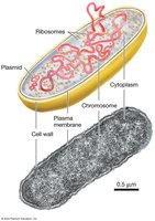
General Structure
Prokaryotic cells are structurally simpler than eukaryotic cells and lack membrane-bound organelles. Their main components include the plasma membrane, cell wall, cytoplasm, ribosomes, and genetic material.
Plasma membrane: Selectively permeable barrier that encloses the cell contents.
Cell wall: Provides structural support and shape.
Cytoplasm: Gel-like substance where cellular processes occur.
Ribosomes: Sites of protein synthesis.
Genetic material: Typically a single, circular chromosome located in the nucleoid region.
Plasmids: Small, circular DNA molecules carrying extra genes, such as those for antibiotic resistance.
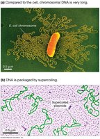
Organization of Prokaryotic DNA
Prokaryotic DNA is organized as a single, circular chromosome. DNA is highly compacted by supercoiling to fit within the cell. Plasmids provide additional genetic capabilities, such as antibiotic resistance, and are important tools in biotechnology.
Nucleoid region: Area where the chromosome is located, not membrane-bound.
Supercoiling: Mechanism for compacting DNA within the cell.
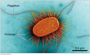
Extracellular Structures
Prokaryotic cells possess specialized extracellular structures for movement and attachment.
Flagella: Long, whip-like structures that rotate to propel the cell through liquid environments.
Fimbriae: Short, hair-like projections that enable attachment to surfaces and play a role in establishing infections.
The Eukaryotic Cell
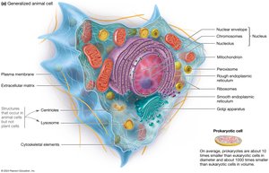
General Animal Cell Structure
Eukaryotic cells are characterized by the presence of membrane-bound organelles, including a nucleus. Animal cells contain various organelles that compartmentalize cellular functions.
Nucleus: Contains genetic material and is surrounded by a nuclear envelope.
Mitochondria: Sites of ATP production via cellular respiration.
Endoplasmic reticulum (ER): Rough ER is studded with ribosomes and synthesizes proteins; smooth ER synthesizes lipids.
Golgi apparatus: Modifies, sorts, and packages proteins and lipids.
Lysosomes: Contain digestive enzymes for breaking down macromolecules.
Peroxisomes: Involved in oxidation reactions and detoxification.
Cytoskeleton: Provides structural support and facilitates movement.
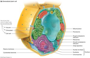
General Plant Cell Structure
Plant cells share many features with animal cells but also have unique structures.
Cell wall: Rigid outer layer composed of cellulose, providing structural support.
Chloroplasts: Sites of photosynthesis, containing chlorophyll.
Central vacuole: Large organelle for storage and maintaining cell turgor.
Plasmodesmata: Channels between plant cells for communication.
Other organelles similar to those in animal cells (nucleus, mitochondria, ER, Golgi, etc.).
Comparison: Prokaryotic vs. Eukaryotic Cells
Prokaryotes: No nucleus, no membrane-bound organelles, generally smaller, single circular chromosome.
Eukaryotes: Nucleus present, many membrane-bound organelles, generally larger, multiple linear chromosomes.
Example: Escherichia coli (prokaryote) vs. human liver cell (eukaryote).
Comparison: Plant vs. Animal Cells
Plant cells: Have cell walls, chloroplasts, and large central vacuoles; perform photosynthesis.
Animal cells: Lack cell walls and chloroplasts; contain centrioles and lysosomes.
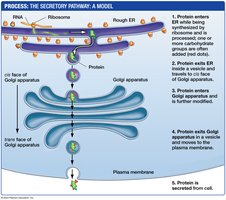
Protein Trafficking and Secretion
The Secretory Pathway
Proteins destined for secretion or for certain organelles follow a specific pathway through the cell, known as the secretory pathway.
Protein synthesis begins on ribosomes in the cytosol.
Proteins with an ER signal sequence are directed to the rough ER, where synthesis is completed and initial modifications occur.
Proteins are packaged into vesicles and transported to the Golgi apparatus for further modification and sorting.
From the Golgi, proteins are sorted and sent to their final destinations: the plasma membrane, lysosomes, or secretion outside the cell.
Targeting Proteins to the ER
Proteins destined for the endomembrane system or secretion possess an ER signal sequence at their N-terminus. This sequence is recognized by the signal recognition particle (SRP), which directs the ribosome to the ER membrane.
Signal peptide: Short amino acid sequence that targets the protein to the ER.
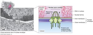
Transport In and Out of the Nucleus
Transport of proteins and RNA between the nucleus and cytoplasm is regulated by the nuclear pore complex (NPC). Proteins destined for the nucleus contain a nuclear localization signal (NLS) that is recognized by import proteins.
Nuclear pore complex: Large protein assembly that spans the nuclear envelope and controls molecular traffic.
Nuclear localization signal (NLS): Amino acid sequence that directs proteins into the nucleus.
Protein Processing in the ER and Golgi Apparatus
In the ER, proteins are folded (often with the help of chaperones) and tagged via glycosylation (addition of carbohydrate groups). The tag determines the protein's final destination. Folded and tagged proteins are sent to the Golgi apparatus for further modification and sorting.
Glycosylation: Addition of carbohydrate groups to proteins, important for sorting and function.
Golgi apparatus: Modifies, sorts, and packages proteins for delivery to their final destinations.
Sorting and Final Destinations from the Golgi
To cell membrane: Default pathway for proteins without a specific carbohydrate tag.
To lysosome: Proteins tagged with mannose-6-phosphate are directed to lysosomes (e.g., acid hydrolase enzymes).
To other organelles: Other carbohydrate tags direct proteins to specific organelles.
Pathways to the Lysosome
Three main pathways deliver material to lysosomes for degradation. Acid hydrolase enzymes, sent from the Golgi, complete lysosome formation and enable breakdown of macromolecules.
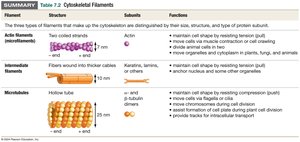
The Cytoskeleton
Overview of Cytoskeletal Filaments
The cytoskeleton is a dynamic network of protein filaments that provides structural support, enables cell movement, and organizes cellular components. There are three main types of cytoskeletal filaments: actin filaments, intermediate filaments, and microtubules.
Filament | Structure | Subunits | Functions |
|---|---|---|---|
Actin filaments (microfilaments) | Two coiled strands, 7 nm diameter | Actin |
|
Intermediate filaments | Fibers wound into thicker cables, 10 nm diameter | Keratin, lamins, or others |
|
Microtubules | Hollow tube, 25 nm diameter | α- and β-tubulin dimers |
|
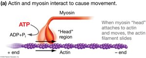
Actin Filaments and Myosin: Movement
Actin filaments interact with the motor protein myosin to produce movement. This interaction is essential for muscle contraction, cell crawling, and cytokinesis (cell division). ATP hydrolysis provides the energy for myosin to "walk" along actin filaments, causing the filaments to slide past each other.
Muscle contraction: Actin and myosin interactions shorten muscle fibers.
Cell crawling: Enables movement of cells such as amoebas and immune cells.
Cytokinesis: Division of the cytoplasm during cell division.
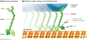
Microtubules and Motor Proteins: Intracellular Transport
Microtubules serve as tracks for the movement of vesicles and organelles within the cell. Motor proteins such as kinesin (moves toward the plus end) and dynein (moves toward the minus end) "walk" along microtubules, powered by ATP hydrolysis. Microtubules are also essential for the movement of cilia and flagella.
Kinesin: Moves cargo toward the cell periphery (plus end of microtubules).
Dynein: Moves cargo toward the cell center (minus end of microtubules).
Flagella and cilia: Structures for cell movement, composed of microtubules arranged in a characteristic pattern.
Summary: Cytoskeletal Filament Functions
Actin filaments: Cell shape, movement, division.
Intermediate filaments: Structural support, anchoring organelles.
Microtubules: Intracellular transport, cell division, movement via cilia/flagella.
Additional info: The cytoskeleton is highly dynamic, allowing cells to rapidly reorganize their internal structure in response to environmental signals or developmental cues.
