 Back
BackMembrane Structure and Function: Study Guide for College Biology
Study Guide - Smart Notes
 Tailored notes based on your materials, expanded with key definitions, examples, and context.
Membrane Structure and Function
Biological Membranes
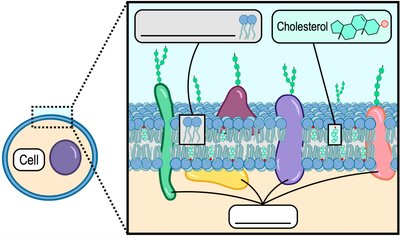
Biological membranes are essential structures that define cell boundaries and regulate the movement of substances in and out of cells. The primary component of these membranes is the phospholipid bilayer, which is amphipathic, meaning it has both hydrophilic and hydrophobic regions. Embedded within this bilayer are proteins and cholesterol, contributing to the membrane's dynamic nature.
Phospholipid Bilayer: Forms the fundamental structure of the membrane, with hydrophilic heads facing outward and hydrophobic tails inward.
Fluid Mosaic Model: Describes the membrane as a fluid structure with a mosaic of proteins embedded within it.
Other Components: Cholesterol stabilizes membrane fluidity; proteins serve various functions.
Synonyms: Also referred to as plasma membrane or cell membrane.
Types of Membrane Proteins
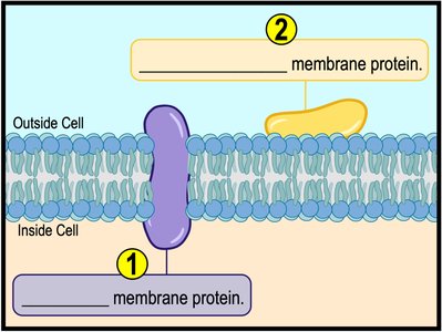
Membrane proteins are crucial for the function and structure of biological membranes. There are two main types:
Integral Membrane Proteins: Span the entire membrane and are embedded within the bilayer.
Peripheral Membrane Proteins: Located on the membrane's perimeter, either inside or outside the cell.
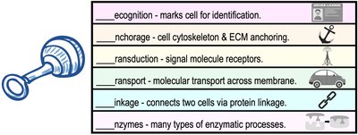
Functions of Membrane Proteins
Recognition: Marks cells for identification.
Anchorage: Anchors cytoskeleton and extracellular matrix.
Transduction: Acts as signal molecule receptors.
Transport: Facilitates molecular transport across the membrane.
Linkage: Connects cells via protein linkage.
Enzymes: Catalyze various enzymatic processes.
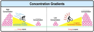
Concentration Gradients and Diffusion
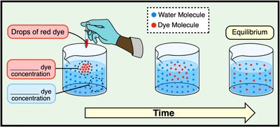
A concentration gradient is the difference in concentration of a substance between two areas. Molecules tend to move down their concentration gradient, from high to low concentration, in a process called diffusion.
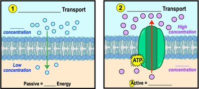
Down Gradient: Movement from high to low concentration (no energy required).
Up Gradient: Movement from low to high concentration (energy required).
Diffusion Example
Diffusion: Movement of dye molecules in water until equilibrium is reached.
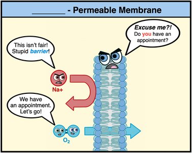
Selective Permeability of Membranes
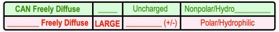
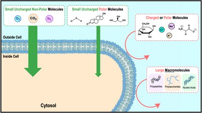
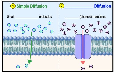
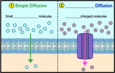
Biological membranes are selectively permeable, meaning they regulate which molecules can cross. Small, uncharged, nonpolar molecules can freely diffuse, while large, charged, or polar molecules require facilitation.
Freely Diffuse: Small, uncharged, nonpolar molecules (e.g., O2, CO2).
Require Facilitation: Large, charged, or polar molecules (e.g., ions, glucose).
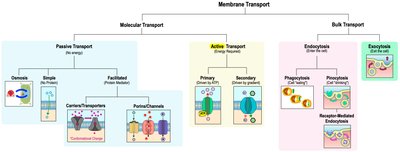
Membrane Transport Overview
Membrane transport is categorized into molecular and bulk transport. Molecular transport includes passive and active mechanisms, while bulk transport involves endocytosis and exocytosis.
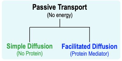
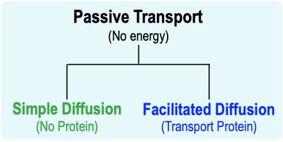
Passive Transport: No energy required; includes osmosis, simple diffusion, and facilitated diffusion.
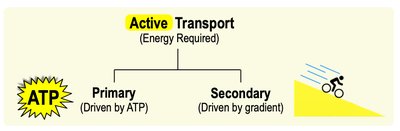
Active Transport: Requires energy (ATP); includes primary and secondary active transport.
Bulk Transport: Endocytosis (phagocytosis, pinocytosis, receptor-mediated) and exocytosis.
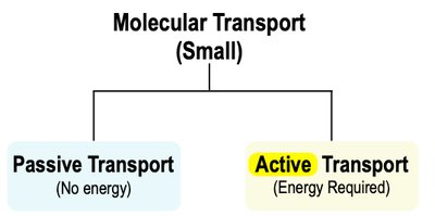
Passive vs. Active Transport
There are two main types of molecular transport across membranes:
Passive Transport: Moves substances down their concentration gradient (high to low), does not require energy.
Active Transport: Moves substances against their concentration gradient (low to high), requires energy (usually ATP).
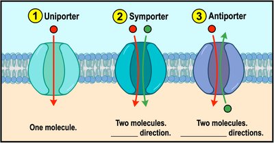
Classes of Membrane Transport Proteins
Uniporters: Transport one molecule at a time in one direction.
Symporters: Cotransport two or more molecules in the same direction.
Antiporters: Cotransport two or more molecules in opposite directions.
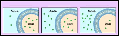
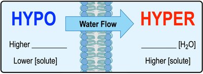
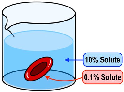
Osmosis
Osmosis is the passive diffusion of water across a semi-permeable membrane. The direction of water flow depends on the relative concentration of solutes in the solutions.
Hypotonic: Lower solute concentration outside the cell; water enters the cell.
Isotonic: Equal solute concentration; water moves in and out at equal rates.
Hypertonic: Higher solute concentration outside the cell; water exits the cell.

Simple and Facilitated Diffusion
Both simple and facilitated diffusion are forms of passive transport, but they differ in their mechanisms:
Simple Diffusion: Direct movement of small, uncharged molecules through the membrane.
Facilitated Diffusion: Movement of charged or polar molecules via transport proteins (channels or carriers).
Transport Proteins in Facilitated Diffusion
Porins/Channels: Form membrane-spanning pores for molecule passage.
Aquaporins: Specialized channels for water transport.
Transporters/Carriers: Undergo conformational changes to move molecules.
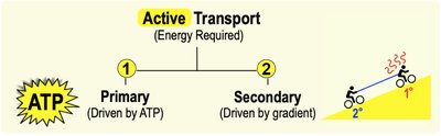
Active Transport
Active transport moves molecules against their concentration gradient and requires energy. There are two types:
Primary Active Transport: Directly uses energy from ATP hydrolysis.
Secondary Active Transport: Uses energy from a concentration gradient established by primary active transport.
Primary Active Transport Example: Na+/K+ Pump
Na+/K+ Pump: Moves sodium ions out and potassium ions into the cell, maintaining essential gradients for cell function.
Uses ATP to transport 3 Na+ out and 2 K+ in per cycle.
Secondary Active Transport Example: Na+-Glucose Cotransporter
Na+ gradient established by primary active transport powers glucose transport against its gradient.
Endocytosis and Exocytosis
Bulk transport mechanisms move large molecules across membranes:
Endocytosis: Cell engulfs macromolecules via vesicles. Types include phagocytosis (cell eating), pinocytosis (cell drinking), and receptor-mediated endocytosis (selective uptake).
Exocytosis: Vesicles fuse with the membrane to release contents outside the cell (e.g., hormones, neurotransmitters).
Summary Table: Membrane Transport Types
Transport Type | Energy Required | Direction | Example |
|---|---|---|---|
Simple Diffusion | No | Down gradient | O2, CO2 |
Facilitated Diffusion | No | Down gradient | Glucose, ions |
Primary Active Transport | Yes (ATP) | Against gradient | Na+/K+ pump |
Secondary Active Transport | Yes (gradient) | Against gradient | Na+-glucose cotransporter |
Endocytosis | Yes | Into cell | Phagocytosis |
Exocytosis | Yes | Out of cell | Hormone secretion |
Key Equations
Diffusion: (Fick's Law: J = flux, D = diffusion coefficient, dC/dx = concentration gradient)
Osmosis: (Osmotic pressure: \Pi = pressure, i = van't Hoff factor, M = molarity, R = gas constant, T = temperature)
Additional info:
Membrane transport is fundamental for cell homeostasis, signaling, and nutrient uptake.
Disruption in transport mechanisms can lead to diseases such as cystic fibrosis or diabetes.
