 Back
BackMicroorganisms and Immunity: Protists, Prokaryotes, and the Immune System
Study Guide - Smart Notes
 Tailored notes based on your materials, expanded with key definitions, examples, and context.
Microorganisms and Immunity
Protists: Diversity and Structure
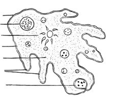
Protists are a diverse group of eukaryotic organisms that are not classified as plants, animals, or fungi. They are primarily aquatic and exhibit a wide range of structures and life strategies.
Definition: Protists are eukaryotes that do not fit into the plant, animal, or fungal kingdoms. They are a polyphyletic group, meaning they do not share a single common ancestor.
Classification: Ongoing debates exist regarding their classification, with some suggesting two kingdoms.
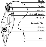
Key Structures:
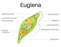
Eyespot: Detects light, aiding producers in finding light and consumers in locating producers.
Contractile Vacuole: Removes excess water, crucial for survival in hypotonic environments.
Pellicle: A protective layer, more rigid than a membrane but less than a cell wall.
Major Protist Examples and Locomotion
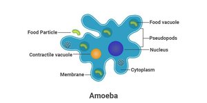
Amoeba: Heterotrophic, shape-shifting protists that move using pseudopods and feed via phagocytosis.
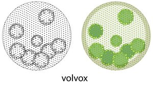
Volvox: Multicellular green algae that move with flagella and reproduce asexually by forming daughter colonies.
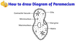
Paramecium: Ciliated protozoans that move and feed using cilia, sweeping food into an oral groove.
Euglena: Flagellated protozoans that may be heterotrophic or autotrophic, possessing chloroplasts and an eyespot.
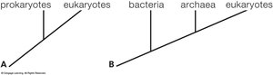
Bacteria and Archaea: Prokaryotic Diversity
Similarities and Differences
Bacteria and Archaea are the two domains of prokaryotes, characterized by the absence of a nucleus and generally unicellular organization.
Shared Features: Both are unicellular, microscopic, and have similar cell structures and shapes.
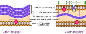
Cell Wall: Most have a cell wall, which prevents bursting in hypotonic environments. The Gram stain technique distinguishes between Gram-positive (thicker peptidoglycan) and Gram-negative (thinner peptidoglycan, outer membrane) bacteria.
Surface Structures: Capsules aid in adhesion; pili and fimbriae assist in movement and attachment.
Reproduction: Prokaryotes reproduce asexually by binary fission, accumulating mutations that drive natural selection.
Gene Transfer: Horizontal gene transfer occurs via transformation, transduction, and conjugation (plasmid exchange).
Endospores: Some bacteria form resistant endospores to survive harsh conditions.
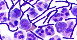
Bacterial Shapes
Bacteria exhibit a variety of shapes, which are important for identification and classification.
Nutrition and Metabolism in Prokaryotes
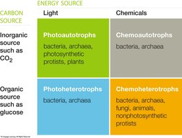
Prokaryotes display diverse metabolic strategies, classified by their energy and carbon sources.
Energy Source | Light | Chemicals |
|---|---|---|
Inorganic Carbon (CO2) | Photoautotrophs (bacteria, archaea, photosynthetic protists, plants) | Chemoautotrophs (bacteria, archaea) |
Organic Carbon (e.g., glucose) | Photoheterotrophs (bacteria, archaea) | Chemoheterotrophs (bacteria, archaea, fungi, animals, nonphotosynthetic protists) |
Biofilms: Surface-coating colonies of prokaryotes that cooperate metabolically and can cause medical and industrial problems.
Bioremediation: Use of prokaryotes to remove pollutants from the environment.
Major Bacterial Lineages
Proteobacteria: All Gram-negative, diverse, some with symbiotic relationships.
Gram-positive Bacteria: Thick cell wall, includes Lactobacillus, Anthrax, Clostridium.
Cyanobacteria: Only prokaryotes with plant-like photosynthesis; some fix nitrogen.
Chlamydias: Intracellular pathogens.
Spirochetes: Helical, motile bacteria; some are pathogenic.
Cell wall thickness | Examples | Gram-stain result | |
|---|---|---|---|
Gram positive | Thicker | Lactobacillus (Yogurt), Anthrax, Food poisoning (Clostridium) |
|
Gram negative | Thinner | E. coli, ulcers (Helicobacter) |
|
Major Archaean Lineages and Habitats
Phylogeny: Archaea are more closely related to eukaryotes than to bacteria.
Habitats: Many live in extreme environments (thermophiles, halophiles, methanogens).
Methanogens: Produce methane from CO2 and H2 (chemoautotrophs):
The Immune System: Defenses Against Pathogens
Overview of Immunity
The immune system protects the body from pathogens through innate and adaptive mechanisms. All animals possess innate immunity, while adaptive immunity is unique to vertebrates.
Innate Immunity: Immediate, non-specific defenses present from birth.
Adaptive Immunity: Specific, acquired defenses that develop after exposure to antigens.
Lines of Immune Defense
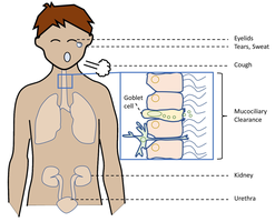
First Line (External Barriers): Physical, chemical, and mechanical barriers prevent pathogen entry (e.g., skin, mucus, lysozyme, stomach acid, cilia, coughing, sneezing).
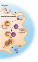
Second Line (Innate Internal Defenses): Activated after pathogens breach barriers; includes phagocytosis, natural killer cells, inflammation, and fever.
Phagocytosis: Neutrophils and macrophages engulf and digest pathogens.
Inflammation: Local response with redness, heat, pain, and swelling.
Fever: Elevated body temperature that inhibits microbes and accelerates repair.
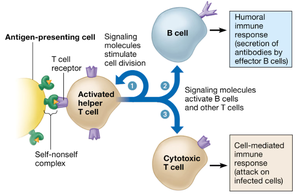
Third Line (Adaptive Immunity): Specific responses involving B and T lymphocytes, memory, and antigen recognition.
Adaptive Immunity: Specific Defenses
Antigens: Molecules that elicit an adaptive immune response (e.g., on pathogens, pollen, transplanted organs).
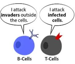
B Lymphocytes (B Cells): Responsible for humoral (antibody-mediated) immunity; produce antibodies against extracellular antigens.
T Lymphocytes (T Cells): Responsible for cell-mediated immunity; attack infected or cancerous cells.
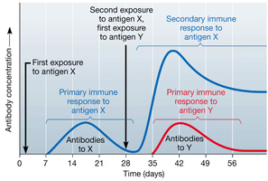
Memory: Adaptive immunity generates memory cells for faster, stronger secondary responses.
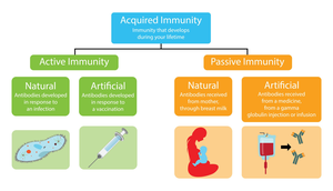
Active vs. Passive Immunity:
Active Immunity: Body produces its own antibodies (infection or vaccination).
Passive Immunity: Antibodies are received from another source (mother, injection).
Humoral (Antibody-Mediated) Immune Response
B cells are activated to produce antibodies specific to antigens found outside cells. These antibodies help neutralize pathogens and toxins and facilitate phagocytosis.
Plasma Cells: Effector B cells that secrete antibodies immediately.
Memory B Cells: Remain in the body for future responses.
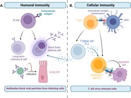
Cell-Mediated Immune Response
T cells are activated to destroy infected or abnormal self cells. Helper T cells coordinate the response by signaling other immune cells, while cytotoxic T cells directly kill infected cells.
Effector T Cells: Act immediately to destroy infected cells.
Memory T Cells: Provide long-term immunity.
Antigen Presentation: Dendritic cells and macrophages present antigens to helper T cells, initiating the adaptive response.
Immune System Disorders
Autoimmune Disorders: Immune system attacks self molecules (e.g., lupus, rheumatoid arthritis, type I diabetes, multiple sclerosis, Crohn’s disease).
Immunodeficiency: Immune system is underactive (e.g., SCID, AIDS, Hodgkin’s disease).
Allergies: Hypersensitive responses to harmless antigens (allergens); mediated by histamine release from mast cells. Severe reactions can cause anaphylactic shock.
