 Back
BackMolecular Biology of the Gene: DNA and RNA Structure, Replication, and Protein Synthesis
Study Guide - Smart Notes
 Tailored notes based on your materials, expanded with key definitions, examples, and context.
DNA: The Molecular Basis of Heredity
Genetic Material and Its Location
Deoxyribonucleic acid (DNA) is the hereditary molecule that stores the instructions for building proteins in all living organisms. In eukaryotic cells, DNA is stored within the nucleus.
DNA contains the genetic code necessary for protein synthesis.
Proteins are essential for cellular structure and function.
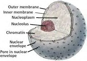
Structure of DNA
DNA is a macromolecule composed of repeating units called nucleotides. Each nucleotide consists of three components:
A phosphate group
A five-carbon sugar (deoxyribose)
A nitrogen-containing base
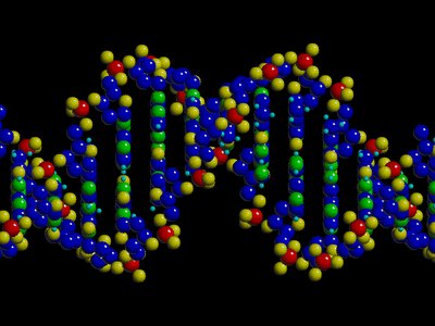
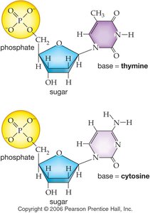
The four nitrogenous bases in DNA are:
Adenine (A)
Thymine (T)
Guanine (G)
Cytosine (C)
Base Pairing and the Double Helix
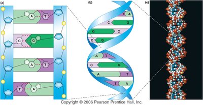
DNA strands are complementary due to specific base pairing:
Adenine (A) pairs with Thymine (T) via two hydrogen bonds.
Guanine (G) pairs with Cytosine (C) via three hydrogen bonds.
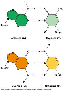
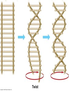
The overall shape of DNA is a double helix, resembling a twisted ladder or spiral staircase. The sugar-phosphate backbone forms the sides, while the nitrogenous bases form the rungs.
Pyrimidines and Purines
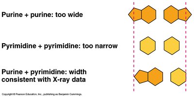
Nitrogenous bases are classified as:
Purines: Double-ring structures (Adenine and Guanine)
Pyrimidines: Single-ring structures (Cytosine and Thymine)
Pairing a purine with a pyrimidine maintains a uniform DNA width of 2 nm.
Genetic Code and Genes
The sequence of bases along DNA encodes genes, which are instructions for building proteins. Each gene corresponds to a specific protein, and the four bases (A, T, C, G) form a genetic alphabet. Triplets of bases (codons) specify amino acids, the building blocks of proteins.
DNA Replication and Repair
Semiconservative Replication
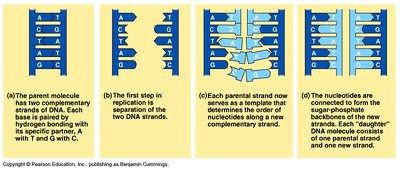
DNA replication is semiconservative: each new DNA molecule consists of one original (parental) strand and one newly synthesized strand. Each strand serves as a template for the other.
Antiparallel Structure
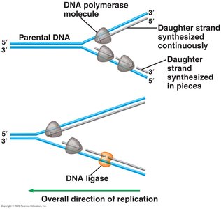
The two DNA strands run in opposite directions (antiparallel): one strand runs 5’ to 3’, the other 3’ to 5’. DNA polymerase synthesizes new DNA only in the 5’ to 3’ direction.
Steps of DNA Replication
Helicase unwinds and unzips the DNA helix, breaking hydrogen bonds between bases.
Single-strand binding proteins stabilize the separated strands, forming a replication fork.
Leading strand: DNA polymerase synthesizes continuously in the 5’ to 3’ direction.
Lagging strand: DNA polymerase synthesizes discontinuously, creating Okazaki fragments, which are later joined by DNA ligase.
Proofreading and Repair
DNA polymerase proofreads new DNA, correcting errors and reducing the mutation rate. Additional repair mechanisms, such as nucleotide excision repair, remove and replace damaged DNA segments.
RNA: Structure and Function
Structure of RNA
Ribonucleic acid (RNA) is a nucleic acid similar to DNA but with key differences:
The sugar is ribose (not deoxyribose).
RNA contains uracil (U) instead of thymine (T).
RNA is usually single-stranded.
Types of RNA
Messenger RNA (mRNA): Carries genetic information from DNA to ribosomes.
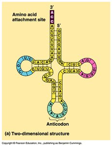
Transfer RNA (tRNA): Brings amino acids to the ribosome during translation; has a cloverleaf structure.
Protein Synthesis: Transcription and Translation
Central Dogma of Molecular Biology
The flow of genetic information follows the central dogma: DNA → RNA → Protein. DNA is transcribed into mRNA, which is then translated into a polypeptide (protein).
Transcription: DNA to mRNA
Initiation: RNA polymerase binds to the promoter region (TATA box) and unwinds DNA.
Elongation: RNA polymerase synthesizes mRNA by adding complementary RNA nucleotides (A pairs with U, G with C).
Termination: RNA polymerase releases the mRNA transcript at the terminator sequence.
Before leaving the nucleus, mRNA is processed: introns (non-coding regions) are removed, exons (coding regions) are spliced together, and a 5’ cap and poly-A tail are added for stability.
Translation: mRNA to Protein
Translation occurs in the cytoplasm and involves:
mRNA: Provides the codon sequence for amino acid assembly.
Ribosomes: Catalyze protein synthesis; composed of large and small subunits.
tRNA: Matches amino acids to codons via its anticodon region.
Ribosomes read mRNA in sets of three nucleotides (codons), each specifying an amino acid. There are 64 possible codons (43), which code for 20 amino acids and include start and stop signals.
Steps of Translation
Small ribosomal subunit binds to mRNA.
Initiator tRNA (with methionine) binds to the start codon (AUG) at the P site.
Large ribosomal subunit attaches, forming the complete ribosome.
tRNA with the next amino acid enters the A site; peptide bond forms between amino acids.
Ribosome shifts, tRNA exits, and the process repeats until a stop codon is reached.
Summary Table: DNA vs. RNA
Feature | DNA | RNA |
|---|---|---|
Sugar | Deoxyribose | Ribose |
Bases | A, T, C, G | A, U, C, G |
Strands | Double-stranded | Single-stranded |
Location | Nucleus (eukaryotes) | Nucleus & Cytoplasm |
Function | Genetic blueprint | Protein synthesis (mRNA, tRNA, rRNA) |
Key Equations and Concepts
Base Pairing Rule: ,
Number of Codons:
Semiconservative Replication: Each daughter DNA = 1 parental + 1 new strand
