 Back
BackNervous System and Nervous Tissue: Structure, Function, and Physiology
Study Guide - Smart Notes
 Tailored notes based on your materials, expanded with key definitions, examples, and context.
Nervous System Overview
Functions of the Nervous System
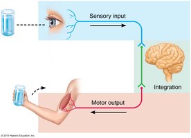
The nervous system is responsible for rapid communication throughout the body, utilizing both electrical and chemical signals. It coordinates sensory input, integration, and motor output to maintain homeostasis and respond to internal and external stimuli.
Sensory Input: Receives information from sensory receptors about changes inside and outside the body.
Integration: Processes and interprets sensory input and determines the appropriate response.
Motor Output: Activates effector organs (muscles and glands) to cause a response.
Organization of the Nervous System
Major Divisions
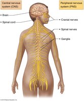
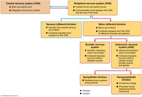
The nervous system is divided into the central nervous system (CNS) and the peripheral nervous system (PNS), each with distinct structures and functions.
Central Nervous System (CNS): Consists of the brain and spinal cord; serves as the integration and control center.
Peripheral Nervous System (PNS): Composed of nerves extending from the CNS (cranial and spinal nerves); connects the CNS to the rest of the body.
Divisions of the Peripheral Nervous System
The PNS is further subdivided based on the direction of information flow and the type of effectors targeted.
Sensory (Afferent) Division: Transmits sensory information from receptors to the CNS.
Somatic Sensory Fibers: Carry information from skin, skeletal muscles, and joints.
Visceral Sensory Fibers: Carry information from visceral organs.
Motor (Efferent) Division: Transmits commands from the CNS to effector organs.
Somatic Nervous System: Controls voluntary movements of skeletal muscles.
Autonomic Nervous System (ANS): Regulates involuntary functions (smooth muscle, cardiac muscle, glands).
Sympathetic Division: Mobilizes body systems during activity ("fight or flight").
Parasympathetic Division: Conserves energy and promotes "rest and digest" functions.
Nervous Tissue
Cell Types
Nervous tissue is composed of two main cell types: neurons and neuroglia (glial cells).
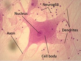
Neurons: Large, highly specialized cells that conduct electrical impulses. They have extreme longevity and a high metabolic rate, requiring continuous oxygen and glucose.
Neuroglia (Glial Cells): Support, protect, and insulate neurons. They are smaller and more numerous than neurons.
Types of Neuroglia
Central Nervous System (CNS)
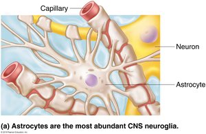
Astrocytes: Support neurons, regulate the extracellular environment, and facilitate nutrient exchange between capillaries and neurons.
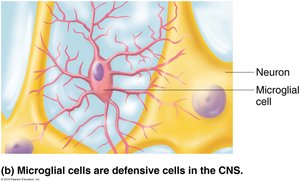
Microglial Cells: Act as immune defense cells, removing debris and pathogens via phagocytosis.
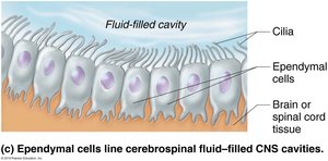
Ependymal Cells: Line cerebrospinal fluid-filled cavities and help circulate cerebrospinal fluid with their cilia.
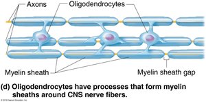
Oligodendrocytes: Produce myelin sheaths that insulate CNS axons and increase signal transmission speed.
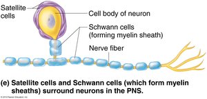
Peripheral Nervous System (PNS)
Satellite Cells: Surround neuron cell bodies in the PNS, providing support and regulating the environment.
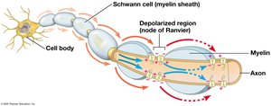
Schwann Cells: Form myelin sheaths around PNS axons, aiding in rapid signal transmission.
Neuron Structure and Classification
Basic Structure of a Neuron
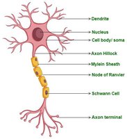
Neurons consist of a cell body (soma) and one or more processes (dendrites and axons). The cell body contains the nucleus and is the biosynthetic center. Dendrites receive signals, while the axon conducts impulses away from the cell body.
Dendrites: Short, branched processes that receive input from other neurons.
Axon: Long process that transmits electrical impulses to other neurons or effectors. The axon hillock is the site of action potential initiation.
Myelin Sheath: Fatty covering that insulates axons and increases the speed of impulse transmission. Gaps in the sheath are called nodes of Ranvier.
Structural Classification of Neurons
Neurons are classified based on the number of processes extending from the cell body:
Type | Description |
|---|---|
Multipolar | Many dendrites, one axon (most common in CNS) |
Bipolar | One dendrite, one axon (rare; found in retina, olfactory mucosa) |
Unipolar (pseudounipolar) | Single process that splits into two branches (sensory neurons in PNS) |
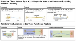
Functional Classification of Neurons
Sensory (Afferent) Neurons: Transmit impulses from sensory receptors to the CNS; mostly unipolar.
Motor (Efferent) Neurons: Carry impulses from the CNS to effectors (muscles/glands); multipolar.
Interneurons: Connect sensory and motor neurons within the CNS; multipolar and most abundant.

Membrane Potential and Action Potentials
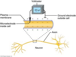
Resting Membrane Potential
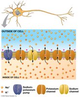
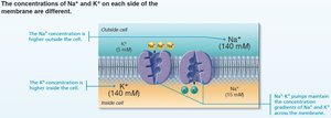
The resting membrane potential is the voltage difference across the neuron's plasma membrane when the cell is not transmitting a signal. It is typically around -70 mV, with the inside of the cell being more negative than the outside.
Maintained by: Sodium-potassium (Na+/K+) pumps and differential permeability of the membrane to Na+ and K+ ions.
Equilibrium Potential: The membrane potential at which the net flow of a particular ion is zero due to a balance between electrical and chemical gradients.
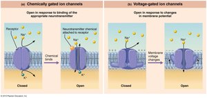
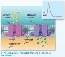
Gated Channels and Ion Movement
Neuronal membranes contain gated ion channels that open or close in response to specific stimuli, allowing selective ion movement and changes in membrane potential.
Chemically Gated Channels: Open in response to neurotransmitter binding.
Voltage-Gated Channels: Open in response to changes in membrane potential.
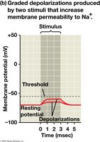
Changes in Membrane Potential
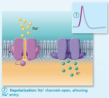
Depolarization: Membrane potential becomes less negative (moves toward zero); increases likelihood of action potential.
Hyperpolarization: Membrane potential becomes more negative; decreases likelihood of action potential.
Graded Potentials: Small, localized changes in membrane potential; magnitude varies with stimulus strength and decays with distance.
Action Potentials: Large, rapid, all-or-nothing depolarizations that propagate along the axon if threshold is reached (typically -55 mV).
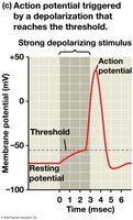
Phases of the Action Potential
Phase | Description |
|---|---|
Resting State | All voltage-gated Na+ and K+ channels are closed; membrane at -70 mV. |
Depolarization | Voltage-gated Na+ channels open; Na+ enters cell, membrane potential rises to +40 mV. |
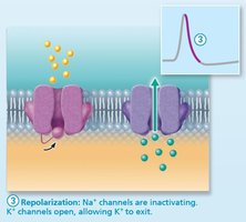
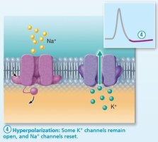
Repolarization | Na+ channels inactivate, K+ channels open; K+ exits cell, membrane potential returns toward -70 mV. |
Hyperpolarization | K+ channels remain open longer, membrane potential drops below resting value before stabilizing. |
Action Potential Conduction
Action potentials propagate along the axon toward synaptic terminals. The refractory period ensures unidirectional flow and limits firing frequency. Myelination and axon diameter affect conduction speed.
Saltatory Conduction: In myelinated axons, action potentials jump between nodes of Ranvier, increasing speed.
Synapses and Neurotransmission
Chemical Synapses
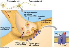
Neurons communicate at synapses, where the presynaptic neuron releases neurotransmitters that bind to receptors on the postsynaptic cell, generating postsynaptic potentials.
Steps:
Action potential arrives at axon terminal.
Voltage-gated Ca2+ channels open; Ca2+ enters presynaptic cell.
Synaptic vesicles fuse with membrane, releasing neurotransmitter into synaptic cleft.
Neurotransmitter binds to ligand-gated ion channels on postsynaptic membrane, altering membrane potential.
Postsynaptic Potentials and Summation
Excitatory Postsynaptic Potential (EPSP): Depolarizes membrane, increasing likelihood of action potential.
Inhibitory Postsynaptic Potential (IPSP): Hyperpolarizes membrane, decreasing likelihood of action potential.
Summation: Multiple EPSPs and IPSPs combine temporally or spatially to influence action potential generation.
Neurotransmitters
Neurotransmitters are chemicals that transmit signals across synapses. Major classes include:
Acetylcholine (ACh): Excitatory at neuromuscular junctions; inhibitory in the heart.
Amino Acids: Glutamate (excitatory), glycine and GABA (inhibitory).
Biogenic Amines: Norepinephrine, epinephrine (fight or flight), dopamine, serotonin (mood, sleep, learning).
Neuropeptides: Chains of amino acids, e.g., endorphins (pain perception).
Practice Questions
Which sequence correctly represents the three main functions of the nervous system?
Answer: Sensory input, integration, motor output
Which division of the nervous system is activated to extend your arm to reach for a beverage?
Answer: Somatic nervous system
Which division is responsible for the rapid heartbeat after a sudden fright?
Answer: Autonomic nervous system
