 Back
BackNeuronal Physiology, Nervous System Organization, and Sensory Systems: Study Guide
Study Guide - Smart Notes
 Tailored notes based on your materials, expanded with key definitions, examples, and context.
Neuronal Physiology
Characteristics of Neurons
Neurons are specialized cells responsible for communication within the nervous system. They possess five major characteristics:
Excitability: Ability to respond to stimuli by initiating electrical changes.
Conductivity: Ability to transmit electrical signals along the cell membrane.
Secretory: Release of neurotransmitters to signal other cells.
Amitotic: Neurons do not undergo mitosis and cannot replicate.
Longevity: Neurons are formed during fetal development and function throughout the lifespan.
Anatomic Structure of a Neuron
The structure of a neuron is closely tied to its function:
Cell Body (Soma): Contains the nucleus and most organelles; site of metabolic activity.
Dendrites: Short, branched processes that receive signals from other neurons.
Axon: Long process that transmits signals away from the cell body; attaches at the axon hillock and ends in synaptic terminals.
Synaptic Terminals: Release neurotransmitters to communicate with postsynaptic cells.
Glial Cells
Glial cells are non-excitable support cells in the nervous system. They are essential for neuron function and exist in similar numbers to neurons.
Astrocytes: Provide structural support and regulate the environment around neurons.
Microglia: Act as immune cells within the CNS.
Ependymal Cells: Line the ventricles of the brain and produce cerebrospinal fluid.
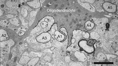
Oligodendrocytes: Form myelin sheaths in the CNS.
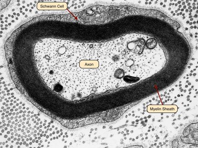
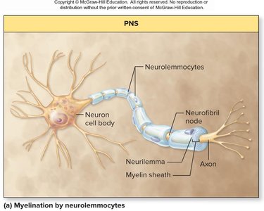
Neurolemmocytes (Schwann Cells): Form myelin sheaths in the PNS.

Myelin and Its Function
Myelin is a multilayered membrane produced by glial cells, insulating axons and increasing the speed of electrical signal transmission.
Oligodendrocytes: Myelinate axons in the CNS.
Schwann Cells: Myelinate axons in the PNS.
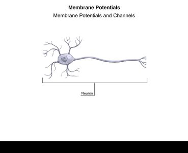
Membrane Potential
All cells have a membrane potential, a voltage difference across the plasma membrane. In neurons, this is crucial for signaling.
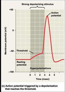
Resting Membrane Potential: The voltage across the membrane when the neuron is not sending signals, typically around -70 mV.
Produced by: The sodium-potassium pump (3 Na+ out, 2 K+ in) and selective permeability of ion channels.
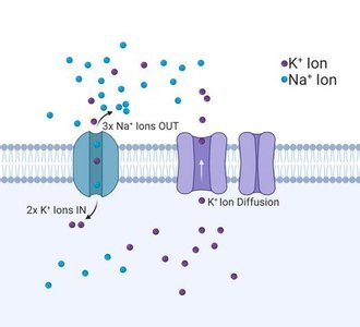
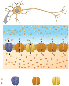
Ion Channels and Neuronal Signaling
Neuronal communication depends on ion channels:
Leak Channels: Always open, allow passive ion movement.
Gated Channels: Open in response to voltage changes or chemical binding.
Voltage-Gated Channels: Open/close in response to changes in membrane potential.
Ligand-Gated Channels: Open/close in response to neurotransmitter binding.
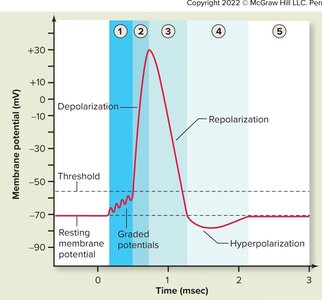
Changes in Membrane Potential
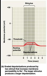
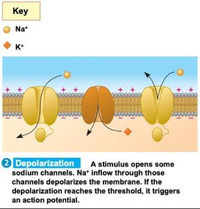
Depolarization: Membrane potential becomes less negative.
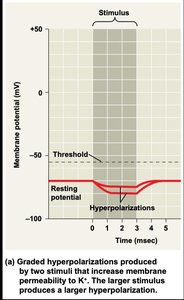
Hyperpolarization: Membrane potential becomes more negative.
Threshold: The critical level of depolarization required to trigger an action potential.
Refractory Period: Time after an action potential when a second cannot be initiated.
Unidirectional Propagation: Action potentials travel in one direction due to refractory period.
Myelinated: Axons covered in myelin, allowing faster signal transmission.
All-or-None: Action potentials occur fully or not at all, regardless of stimulus strength.
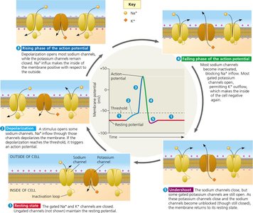
Action Potential: Steps and Recovery
An action potential is a rapid change in membrane potential that propagates along the axon.
Resting Potential: Neuron is at rest; most voltage-gated channels closed.
Threshold: Sufficient depolarization opens some Na+ channels.
Rising Phase: Massive influx of Na+ causes rapid depolarization.
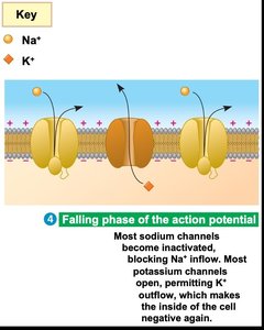
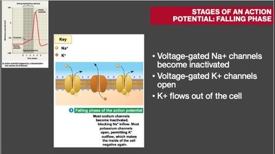
Falling Phase: Na+ channels inactivate, K+ channels open, K+ exits cell.
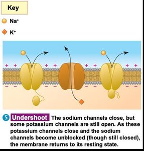
Undershoot: K+ channels stay open too long, membrane potential falls below resting.
Recovery: Na+/K+ pump restores resting potential.


Refractory Period and Unidirectional Propagation
Ensures action potentials travel in one direction, from axon hillock to synaptic terminals.
Limits the frequency of action potentials.
All-or-None Law
Action potentials are always the same size and intensity once threshold is reached.
Stimulus strength affects frequency, not amplitude.
Adaptations for Speed of Conduction
Increased Axon Diameter: Larger diameter allows faster signal transmission (e.g., squid giant axon).
Myelination: Myelin sheath enables saltatory conduction, where action potentials jump between nodes of Ranvier, increasing speed and efficiency.
Organization of Nervous Systems
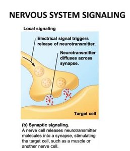
Neuronal Communication: Chemical Synapse
Neurons communicate via chemical synapses, where neurotransmitters transmit signals between cells.
Presynaptic neuron synthesizes and packages neurotransmitter in vesicles.
Arrival of action potential causes release of neurotransmitter.
Neurotransmitter diffuses across synaptic cleft and binds to ligand-gated ion channels on postsynaptic cell.
Binding triggers a response in the postsynaptic neuron.
Nervous System Organization in Animals
Diffuse Nervous System: Simple nerve net, no centralization (e.g., jellyfish).
Centralized Nervous System: Collection of neurons in a central location (e.g., brain), allows greater integration and processing.
Nerve: Bundle of axons from multiple neurons.
Vertebrate Nervous System: CNS and PNS
Central Nervous System (CNS): Brain and spinal cord; site of integration, processing, and myelination by oligodendrocytes.
Peripheral Nervous System (PNS): Sensory detection and motor actions throughout the body; myelination by Schwann cells.
Functional Hierarchy of the Vertebrate Nervous System
Afferent Division: Sensory input toward CNS.
Efferent Division: Motor output away from CNS.
Somatic: Voluntary control.
Visceral: Relates to organs.
Autonomic: Involuntary control.
Autonomic Nervous System
Sympathetic: Fight or flight responses.
Parasympathetic: Rest and digest responses.
Enteric: Controls digestive tract, pancreas, and gallbladder.
Specialized Regions of the CNS
Spinal Cord: Conveys information, generates locomotion patterns, produces reflexes.
Gray Matter: Neuron cell bodies.
White Matter: Myelinated axons.
Brain Divisions: Forebrain (cognition), midbrain (relay), hindbrain (movement, balance).
Cerebrum: Higher cognitive functions, divided into four lobes: frontal (motor, personality), parietal (touch), temporal (hearing, smell), occipital (vision).
Sensory Systems
Stages of Sensory Signal Processing
Organisms sense their environment through a general pathway:
Sensory Reception: Sensory receptor detects a stimulus.
Signal Transduction: Conversion of stimulus energy into a change in membrane potential.
Transmission: Information sent as action potentials.
Modulation: Amplification or adaptation of the signal.
Perception: Brain interprets electrical signals as sensory stimuli.
Encoding Information
Action potential size is constant; frequency encodes stimulus intensity.
Refractory period limits maximum firing rate.
Major Sensory Modalities and Receptors
Mechanoreceptors: Detect mechanical force (pressure, touch, stretch, motion).
Electromagnetic Receptors: Detect light, electricity, magnetism.
Thermoreceptors: Detect temperature changes.
Chemoreceptors: Detect chemicals (smell, taste).
Pain Receptors (Nociceptors): Detect noxious stimuli and tissue damage.
Summary Table: Sensory Modalities and Receptors
Modality | Receptor Type | Example |
|---|---|---|
Touch | Mechanoreceptor | Whiskers in cats |
Vision | Electromagnetic receptor | Photoreceptors in eyes |
Temperature | Thermoreceptor | Hypothalamus in humans |
Taste/Smell | Chemoreceptor | Olfactory cells |
Pain | Nociceptor | Withdrawal reflex |
Additional info: Sensory adaptation allows organisms to ignore constant stimuli, while amplification strengthens weak signals during transduction.
