 Back
BackSkeletal, Muscular, and Nervous Systems: Structure and Function
Study Guide - Smart Notes
 Tailored notes based on your materials, expanded with key definitions, examples, and context.
Chapter 11 – Skeletal System and Motion
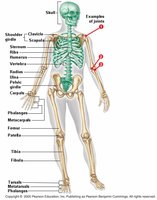
The Vertebrate Skeleton
The vertebrate skeleton provides structural support, protection for internal organs, and facilitates movement. It is divided into two main parts: the axial skeleton and the appendicular skeleton.
Axial skeleton: Includes the cranium (skull) and vertebral column (spine).
Appendicular skeleton: Comprises the pectoral and pelvic girdles, as well as the bones of the arms and legs.
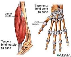
The Skeleton: Bones and Joints
Bones are living organs that come together at joints, allowing movement. Cartilage, ligaments, and tendons are essential connective tissues associated with bones and joints.
Cartilage: Cushions joints, prevents bones from rubbing together, and acts as a shock absorber.
Ligaments: Strong fibrous tissues that connect bone to bone, stabilizing joints.
Tendons: Connect muscle to bone, transmitting the force needed for movement.
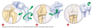
The Human Skeleton: Articulation and Joint Types
The versatility of the vertebrate skeleton is due to its movable joints, which allow for a wide range of motion. Different types of joints permit different movements:
Ball-and-socket joints: Allow movement in all directions (e.g., shoulder, hip).
Hinge joints: Permit movement in one plane (e.g., knee, elbow).
Pivot joints: Allow bones to rotate (e.g., between the radius and ulna in the forearm).
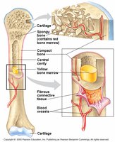
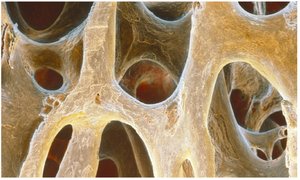
Bone Structure and Remodeling
Bones are complex organs composed of living tissues. The outer surface is covered by fibrous connective tissue, which aids in bone repair. The interior contains a matrix of collagen fibers and minerals, providing both flexibility and strength.
Bone matrix: Made of flexible collagen fibers embedded in hard calcium and phosphate.
Bone remodeling: Specialized cells build new bone and reabsorb minerals as needed for body function.
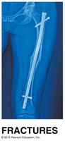
Skeletal System Disorders
The skeletal system can be affected by various disorders:
Fractures: Occur when forces exceed a bone’s ability to flex, resulting in a break.
Arthritis: Inflammation of the joints, caused by aging, immune disorders, injury, or infection.
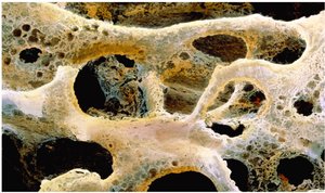
Osteoporosis: Impaired bone remodeling leads to porous, fragile bones and increased fracture risk.
Condition | Main Feature |
|---|---|
Fracture | Break in bone continuity |
Arthritis | Joint inflammation |
Osteoporosis | Porous, weak bones |
Muscular System
Muscle Structure and Function
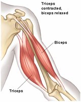
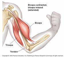
The muscular system is responsible for movement. Skeletal muscles are attached to bones and work in antagonistic pairs to produce motion.
Origin: The fixed attachment point of a muscle.
Insertion: The movable attachment at a joint.
Antagonistic pairs: While one muscle contracts, its pair relaxes (e.g., biceps and triceps).
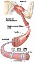
Muscle Cells and Contraction
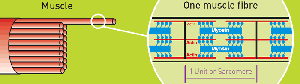
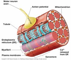
Muscle cells, or fibers, are organized into bundles. Each fiber contains myofibrils, which are further divided into sarcomeres—the functional units of contraction.
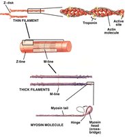
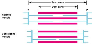
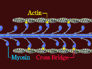
Sarcomere: Composed of overlapping thick (myosin) and thin (actin) filaments.
Contraction: Occurs when thin filaments slide past thick filaments, shortening the sarcomere.
Mechanism of Muscular Contraction: Sliding Filament Model
The sliding filament model explains how muscles contract. Myosin heads bind to actin, using ATP to power the movement that slides filaments past each other.
ATP: Provides energy for myosin head movement.
Power stroke: Myosin pulls actin toward the center of the sarcomere, shortening the muscle.
Regulation: Calcium ions and regulatory proteins control access to binding sites.
Controlling Muscular Contraction
Muscle contraction is controlled by nerve impulses, calcium ions, and ATP. An action potential triggers the release of calcium from the sarcoplasmic reticulum, enabling contraction. Removal of calcium stops contraction.
Action potential: Electrical signal from a motor neuron.
Calcium: Binds to regulatory proteins, exposing myosin binding sites on actin.
ATP: Required for both contraction and relaxation.
Types of Muscle Tissue
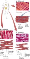
There are three main types of muscle tissue in the body:
Skeletal muscle: Voluntary, striated muscle attached to bones.
Cardiac muscle: Involuntary, striated muscle found in the heart.
Smooth muscle: Involuntary, non-striated muscle found in internal organs.
Chapter 11 – Nervous System
Organization of the Nervous System
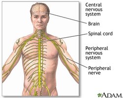
The nervous system is the body's communication network, divided into the central nervous system (CNS) and peripheral nervous system (PNS).
CNS: Consists of the brain and spinal cord; processes and integrates information.
PNS: Composed of nerves that connect the CNS to the rest of the body.
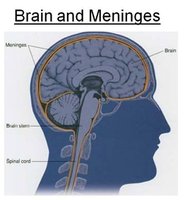
Central Nervous System: Structure and Protection
The CNS is protected by the meninges (connective tissue layers) and cushioned by cerebrospinal fluid, which also supplies nutrients and removes waste.
Brain: Integrates sensory information, controls muscles, and is the center of intellect and emotion.
Spinal cord: Main communication pathway between brain and body.
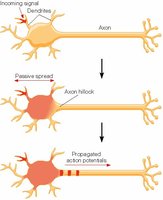
Structure and Function of Nerve Cells
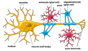
Neurons are specialized cells that transmit electrical and chemical signals. Glial cells support and nourish neurons.
Dendrites: Receive information from other neurons.
Cell body: Integrates incoming signals.
Axon: Transmits signals to other neurons or effectors.
Synapse: The junction between neurons where communication occurs.
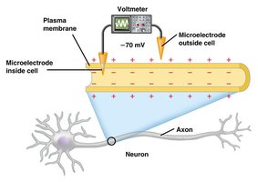
Resting Membrane Potential
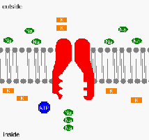
At rest, a neuron has a charge difference across its membrane, with the inside being more negative than the outside. This is maintained by ion gradients and selective membrane permeability.
Na+: Higher concentration outside the cell.
K+: Higher concentration inside the cell.
Sodium-potassium pump: Uses ATP to maintain ion gradients.
Action Potential and Nerve Signal Propagation
An action potential is a rapid change in membrane potential that travels along the axon, allowing communication over long distances.
Depolarization: Na+ channels open, Na+ enters the cell, making the inside more positive.
Repolarization: K+ channels open, K+ exits, restoring negativity inside.
Propagation: The action potential regenerates along the axon, similar to a wave or falling dominoes.
Myelination: Glial cells insulate axons, allowing the impulse to jump between nodes (saltatory conduction), increasing speed.
Chemical Synapses and Neurotransmitters
At synapses, electrical signals are converted to chemical signals via neurotransmitters, which cross the synaptic cleft to transmit the impulse to the next cell.
Neurotransmitters: Chemicals such as dopamine, acetylcholine, and norepinephrine transmit signals across synapses.
Synaptic vesicles: Store neurotransmitters in the presynaptic neuron.
Synaptic cleft: The gap between neurons where neurotransmitters are released.
Neurotransmitter | Main Function |
|---|---|
Dopamine | Brain signaling; lack associated with Parkinson’s disease |
Acetylcholine | Nerve-to-muscle signaling |
Norepinephrine | Mood and behavior regulation |
Nervous System Disorders
The nervous system can be affected by various diseases and injuries, which are often difficult to diagnose and treat due to system complexity.
Examples: Depression, Alzheimer’s disease, paralysis, spinal infections.
