 Back
BackStructure and Function of Cells: An Introduction to Cell Biology
Study Guide - Smart Notes
 Tailored notes based on your materials, expanded with key definitions, examples, and context.
Introduction to Cells & Their Structures
Overview
Cells are the fundamental units of life, forming the basis of all living organisms. This section introduces the cell theory, the classification of cells, and the structural and functional diversity found within cells. Understanding these concepts is essential for further study in biology and human physiology.
Cell Theory
Principles of Cell Theory
All living things are composed of cells and cell products.
The cell is the smallest unit that exhibits all the characteristics of life.
All cells arise only from preexisting cells.
These principles, established through centuries of observation and experimentation, remain foundational in biology today.
Classification of Cells
Prokaryotic vs. Eukaryotic Cells
Cells are classified based on their internal organization and the presence or absence of membrane-bound organelles.
Prokaryotic Cells: Simpler structure, lack a true nucleus, DNA is located in a nucleoid region, no membrane-bound organelles, typically have a rigid cell wall. Examples: Bacteria, blue-green algae.
Eukaryotic Cells: More complex, possess a true nucleus surrounded by a nuclear membrane, contain various membrane-bound organelles, cytoplasm filled with cytosol. Examples: Animals, plants.
Internal organization is the key determinant of cell classification.
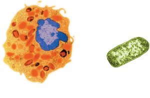
Figure: Comparison of eukaryotic (left) and prokaryotic (right) cell structures. Eukaryotic cells have a prominent nucleus and organelles, while prokaryotic cells lack these features.
Structure Reflects Function
Specialization of Cells
Although all cells share basic functions such as gathering raw materials, excreting wastes, synthesizing macromolecules, and reproducing, their structures are specialized to support specific roles:
Muscle cells: Abundant organelles for energy production.
Nerve cells: Long, thin structures for signal transmission.
Red blood cells: Round and flexible to traverse capillaries.
Every eukaryotic cell exhibits some degree of functional specialization.
Cell Size and Efficiency
Surface Area to Volume Ratio
Cells remain small to maximize efficiency in exchanging materials with their environment. As a cell grows, its volume increases faster than its surface area, limiting the rate at which materials can cross the plasma membrane.
Smaller cells: More efficient exchange of materials.
Microvilli: Surface projections that increase surface area, common in digestive tract and kidney tubules.
An eightfold increase in cell volume results in only a fourfold increase in surface area, emphasizing the importance of small cell size.
Microscopes in Cell Biology
Types of Microscopes
Microscopes are essential tools for visualizing cells and their structures:
Light Microscope: Magnifies up to 1000x, suitable for general cell observation.
Transmission Electron Microscope (TEM): Provides 2D images of internal structures, magnifies up to 100,000x.
Scanning Electron Microscope (SEM): Produces 3D images of cell surfaces, magnifies up to 100,000x.
Internal Cell Structures & Functions
Membrane-Bound Organelles
Eukaryotic cells contain specialized structures called organelles, each responsible for distinct cellular functions.
The Nucleus
Structure and Function
The nucleus serves as the control center of the cell, housing most of the genetic material (DNA).
Nuclear membrane: Double phospholipid bilayer that encloses DNA.
Nucleolus: Dense region where ribosomal components are formed.
Nuclear pores: Allow selective movement of materials (e.g., ribosomes, proteins, RNA) in and out of the nucleus, but are too small for DNA to exit.
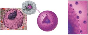
Figure: Transmission electron micrograph and diagram of the nucleus, highlighting the nucleolus and nuclear pores.
Ribosomes
Protein Synthesis
Ribosomes are assembled from RNA and proteins in the nucleolus, then transported to the cytoplasm. They may float freely or attach to the endoplasmic reticulum. Ribosomes are the sites of protein synthesis, assembling amino acids into polypeptide chains according to genetic instructions.
Endomembrane System
Components and Functions
The endomembrane system includes the rough and smooth endoplasmic reticulum, Golgi apparatus, and specialized vesicles. These structures coordinate the synthesis, modification, packaging, and transport of cellular products.
Endoplasmic Reticulum (ER)
Rough ER: Studded with ribosomes, involved in protein synthesis and initial folding.
Smooth ER: Lacks ribosomes, synthesizes lipids and hormones, packages proteins and lipids for transport to the Golgi apparatus.
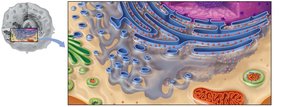
Figure: Diagram showing the rough and smooth ER, with ribosomes attached to the rough ER and vesicles budding off for transport.
Golgi Apparatus
The Golgi apparatus receives vesicles from the ER, refines and sorts proteins and lipids, adds molecular labels, and packages them for delivery to their final destinations. Lipid synthesis also occurs here.
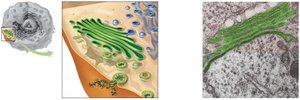
Figure: The Golgi apparatus receives, modifies, and ships cellular products via vesicles.
Vesicles
Secretory vesicles: Export products out of the cell.
Endocytotic vesicles: Bring external materials into the cell.
Peroxisomes: Contain enzymes to break down toxic wastes.
Lysosomes: Digest bacteria, large particles, and perform cellular housekeeping.
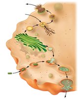
Figure: Pathways of vesicle formation and fusion, showing the roles of lysosomes and peroxisomes in cellular digestion and detoxification.
Mitochondria
Energy Production
Mitochondria are the powerhouses of the cell, converting nutrients into ATP through cellular respiration. They have a double membrane, with the inner membrane folded into cristae to increase surface area for energy-releasing reactions. Mitochondria contain their own DNA and ribosomes, reflecting their evolutionary origins.
Cytoskeleton
Support and Structure
The cytoskeleton is a network of microtubules (hollow tubes) and microfilaments (solid fibers) that provides structural support, maintains cell shape, and anchors organelles. It also facilitates intracellular transport and cell movement.
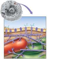
Figure: The cytoskeleton connects to the plasma membrane and supports cellular architecture.
Cilia and Flagella
Cellular Movement
Cilia: Short, numerous projections made of microtubules; move materials along cell surfaces (e.g., in respiratory tract).
Flagella: Longer, fewer in number; propel cells (e.g., sperm cells).
Centrioles
Role in Cell Division
Centrioles are short, rod-like structures near the nucleus, essential for organizing microtubules during cell division. They ensure proper alignment and separation of genetic material.
Summary Table: Major Eukaryotic Cell Structures and Functions
Structure | Main Function |
|---|---|
Plasma membrane | Controls movement of materials into and out of cell |
Nucleus | Information center; contains DNA |
Ribosomes | Site of protein synthesis |
Rough ER | Protein synthesis and processing |
Smooth ER | Lipid and hormone synthesis; packaging |
Golgi apparatus | Refines, packages, and ships macromolecules |
Lysosome | Digests damaged organelles and debris |
Peroxisome | Destroys toxic waste |
Mitochondrion | Produces energy (ATP) |
Cytoskeleton | Structural framework |
Centrioles | Involved in cell division |
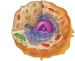
Figure: Diagram of a eukaryotic cell showing the location and function of major organelles.
References: Johnson, M.D. (2017). Human biology: Concepts and current issues (8th ed). Pearson Education Inc. Johnson, M.D. & Long, S (2021). Human biology: Concepts and current issues (9th ed). Pearson Education Inc.
