 Back
BackStructure and Function of Cells: Cell Theory, Types, and Organelles
Study Guide - Smart Notes
 Tailored notes based on your materials, expanded with key definitions, examples, and context.
Cell Theory and Classification
Cell Theory
The cell theory is a foundational concept in biology, describing the properties and significance of cells in living organisms.
All living things are composed of cells and cell products.
The cell is the smallest unit that exhibits all the characteristics of life.
All cells arise only from preexisting cells.
These principles remain central to modern biology and guide our understanding of life at the cellular level.
Classification of Cells
Cells are classified based on their internal organization into two main types: prokaryotic and eukaryotic cells.
Prokaryotic Cells: Simpler structure, lack membrane-bound organelles, DNA is located in a nucleoid region, surrounded by a plasma membrane and often a rigid cell wall. Examples: Bacteria, blue-green algae.
Eukaryotic Cells: More complex, contain a true nucleus (membrane-bound DNA), cytoplasm with specialized organelles, surrounded by a plasma membrane. Examples: Animals, plants.
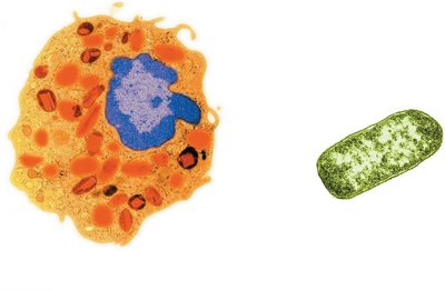
Example: Animal cells (eukaryotic) have a large nucleus and many organelles, while bacterial cells (prokaryotic) lack these features.
Cell Structure and Function
Structure Reflects Function
Although all cells share basic functions—such as gathering raw materials, excreting wastes, synthesizing macromolecules, and reproducing—their structures are specialized for their roles.
Muscle cells: Abundant organelles for energy production.
Nerve cells: Long and thin to transmit signals over distances.
Red blood cells: Round and flexible to move through capillaries.
Each eukaryotic cell type is specialized for its function.
Cell Size and Efficiency
Cells remain small to maximize efficiency in exchanging materials with their environment. As a cell grows, its volume increases faster than its surface area, limiting the rate of material exchange.
Metabolic activities are proportional to cell volume.
Material exchange occurs across the plasma membrane (surface area).
Microvilli are microscopic projections that increase surface area, common in digestive tract and kidney tubules.
Example: Cells with microvilli can absorb nutrients more efficiently due to increased surface area.
Microscopy
Types of Microscopes
Microscopes are essential for studying cells due to their small size. The main types include:
Light Microscope: Magnifies up to 1000x, suitable for viewing live cells and general structures.
Transmission Electron Microscope (TEM): Provides 2D images of internal structures, magnifies up to 100,000x.
Scanning Electron Microscope (SEM): Produces 3D images of cell surfaces, magnifies up to 100,000x.
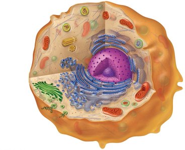
Internal Cell Structures (Organelles)
The Nucleus
The nucleus is the control center of eukaryotic cells, containing most of the genetic material (DNA).
Nuclear membrane: Double phospholipid layer that encloses DNA.
Nucleolus: Dense region where ribosome components are formed.
Nuclear pores: Allow selective movement of materials (e.g., ribosomes, proteins, RNA) in and out of the nucleus; DNA remains inside.
Ribosomes
Ribosomes are the sites of protein synthesis, assembling amino acids into specific protein chains.
Formed from RNA and proteins in the nucleolus.
Can be free in the cytoplasm or attached to the endoplasmic reticulum.
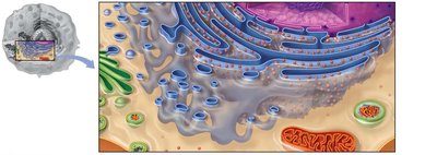
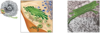
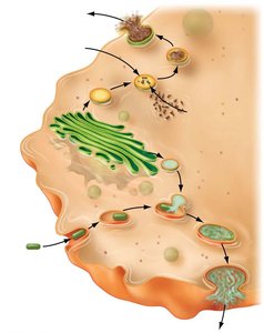
Endomembrane System
The endomembrane system includes several interconnected organelles responsible for synthesis, packaging, and transport of cellular materials.
Rough Endoplasmic Reticulum (RER): Studded with ribosomes, synthesizes proteins.
Smooth Endoplasmic Reticulum (SER): Lacks ribosomes, synthesizes lipids and some hormones, packages proteins and lipids for transport.
Golgi Apparatus: Receives, refines, sorts, and ships products from the ER; also involved in lipid synthesis.
Specialized Vesicles: Transport and store materials within the cell.
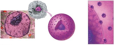
Vesicles
Vesicles are small, membrane-bound sacs that transport and store substances within cells.
Secretory vesicles: Export products out of the cell.
Endocytotic vesicles: Bring external materials into the cell.
Peroxisomes: Contain enzymes to destroy toxic wastes.
Lysosomes: Contain digestive enzymes to break down bacteria, large particles, and cellular debris.
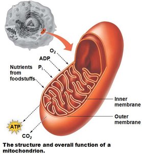
Mitochondria
Mitochondria are the powerhouses of the cell, converting nutrients into ATP through cellular respiration.
Double membrane: Outer membrane is smooth; inner membrane is folded (cristae) and contains enzymes for energy production.
Own DNA and ribosomes: Reflects evolutionary origins.
Number varies: Cells with higher energy demands (e.g., muscle cells) have more mitochondria.
Equation for Cellular Respiration:
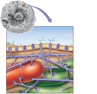
Cytoskeleton
The cytoskeleton provides structural support, maintains cell shape, and anchors organelles.
Microtubules: Hollow tubes for support and transport.
Microfilaments: Thin fibers for movement and shape changes.
Connect to glycoproteins in the plasma membrane.
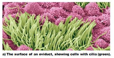
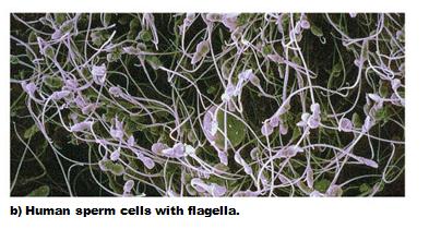
Cilia and Flagella
Cilia and flagella are structures made of microtubules that enable movement.
Cilia: Short, numerous, move materials along cell surfaces (e.g., in the respiratory tract, oviduct).
Flagella: Long, few in number, move the entire cell (e.g., sperm cells).
Centrioles
Centrioles are short, rod-like structures near the nucleus, essential for cell division. They help align and separate genetic material during mitosis and meiosis.
Summary Table: Major Eukaryotic Cell Structures and Functions
Structure | Main Function |
|---|---|
Plasma membrane | Controls movement of materials into and out of cell |
Nucleus | Information center; contains DNA |
Ribosomes | Site of protein synthesis |
Rough ER | Protein synthesis by ribosomes |
Smooth ER | Macromolecule synthesis (other than proteins) |
Golgi apparatus | Refines, packages, and ships macromolecular products |
Lysosome | Digests damaged organelles and debris |
Peroxisome | Destroys cellular toxic waste |
Mitochondrion | Produces energy (ATP) for the cell |
Cytoskeleton | Structural framework of the cell |
Centrioles | Involved in cell division |
Secretory vesicle | Membrane-bound shipping container |
Additional info: The above notes integrate and expand upon the provided lecture content, ensuring a comprehensive overview suitable for college-level biology students.
