 Back
BackStructure and Function of the Plasma Membrane
Study Guide - Smart Notes
 Tailored notes based on your materials, expanded with key definitions, examples, and context.
Plasma Membrane: Structure and Function
Overview of the Plasma Membrane
The plasma membrane is the outermost boundary of a cell, maintaining the cell as a distinct unit and providing organized metabolic areas. It is approximately 3.5 nanometers thick and is primarily composed of a phospholipid bilayer. The plasma membrane allows interaction between the cell's internal (intracellular) and external (extracellular) environments and is essential for maintaining homeostasis.
Phospholipids: Molecules with a polar (hydrophilic) head and two non-polar (hydrophobic) tails, forming the bilayer structure.
Cholesterol: Increases the strength and rigidity of the membrane.
Proteins: Embedded within the membrane, these assist with the movement of materials and communication between cells.
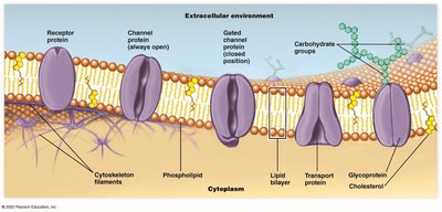
Embedded Proteins in the Plasma Membrane
Proteins embedded in the plasma membrane perform a variety of functions, including transport, signaling, and cell recognition.
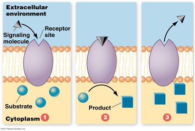
Receptor Proteins: Span the membrane and transmit information by binding signaling molecules, triggering chemical reactions inside the cell.
Channel Proteins: Form open or gated channels for the movement of ions and water.
Transport Proteins: Change shape to move specific molecules across the membrane.
Glycoproteins: Proteins with attached carbohydrate groups, important for cell recognition and communication.
Movement Across the Plasma Membrane
Passive Transport
Passive transport does not require energy and relies on the natural movement of molecules from areas of high concentration to low concentration (down their concentration gradient).
Diffusion: Movement of molecules (e.g., O2, CO2, urea) directly through the lipid bilayer.
Diffusion Through Channels: Water and ions (e.g., Na+, K+, Ca2+) move through protein channels, which may be always open or gated.
Facilitated Diffusion: Specific molecules (e.g., glucose) bind to carrier proteins, which change shape to transport them across the membrane. This process is highly selective.
Osmosis: The diffusion of water across a selectively permeable membrane, moving from areas of high water concentration to low water concentration.
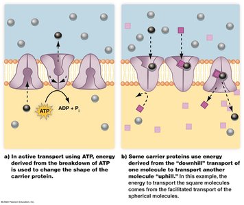
Active Transport
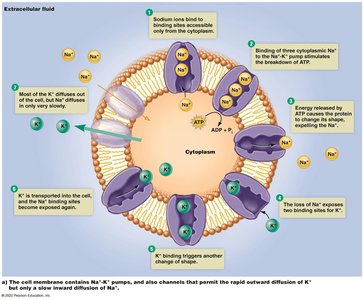
Active transport moves substances against their concentration gradient (from low to high concentration) and requires energy, usually from ATP. Membrane proteins act as pumps to move molecules in or out of the cell.
ATP-driven pumps: Use energy from ATP hydrolysis to change the shape of the carrier protein and transport molecules.
Coupled transport: The energy from passive transport of one molecule is used to power the active transport of another.
Example: The sodium-potassium (Na+/K+) pump is a classic example of active transport in animal cells.
Equation for ATP hydrolysis:
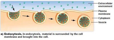
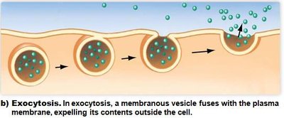
Bulk Transport: Endocytosis and Exocytosis
Bulk transport mechanisms move large molecules or large quantities of substances across the plasma membrane.
Endocytosis: The cell engulfs material from the extracellular environment, forming a vesicle that brings the material into the cell. This process can be selective or non-selective.
Exocytosis: Vesicles formed within the cell fuse with the plasma membrane, releasing their contents to the extracellular environment. This is important for secretion of products and removal of wastes.
Tonicity and Its Effects on Cells
Definition and Importance of Tonicity
Tonicity refers to the relative concentration of solutes in the fluid inside and outside the cell. It determines the direction of water movement across the plasma membrane and is crucial for maintaining cell volume and function.
Isotonic Solution: Solute concentrations are equal inside and outside the cell; water movement is balanced, and cell volume remains stable.
Hypertonic Solution: Higher solute concentration outside the cell; water moves out, causing the cell to shrink (crenation).
Hypotonic Solution: Higher solute concentration inside the cell; water moves in, causing the cell to swell and potentially burst (lysis).
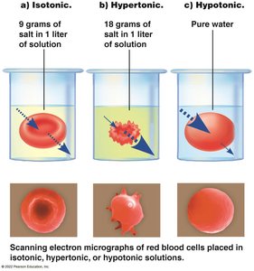
Example: Red blood cells placed in different solutions will change shape depending on the tonicity of the surrounding fluid.
Summary Table: Effects of Tonicity on Cells
Solution Type | Solute Concentration | Water Movement | Effect on Cell |
|---|---|---|---|
Isotonic | Equal inside and outside | No net movement | Cell remains normal |
Hypertonic | Higher outside | Water moves out | Cell shrinks (crenation) |
Hypotonic | Higher inside | Water moves in | Cell swells and may burst (lysis) |
Additional info: Understanding membrane transport is essential for topics such as nerve impulse transmission, nutrient absorption, and kidney function in human biology.
