 Back
BackStudy Guide: Structure and Function of Large Biological Molecules, Cell Structure, and Membrane Function
Study Guide - Smart Notes
 Tailored notes based on your materials, expanded with key definitions, examples, and context.
Chapter 5: The Structure and Function of Large Biological Molecules
Monomers and Polymers
Biological macromolecules are composed of smaller units called monomers, which join together to form polymers. These polymers include carbohydrates, proteins, and nucleic acids, each serving essential functions such as storage, structure, and information transfer.
Monomer: A small molecule used repetitively to build polymers.
Polymer: A chain of repeating monomer units.
Examples: Carbohydrates (e.g., glucose), proteins (e.g., amino acids), nucleic acids (e.g., nucleotides).
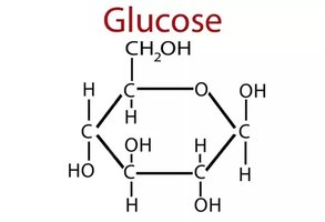
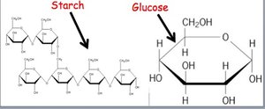
Polysaccharides: Structure and Function
Polysaccharides are complex carbohydrates formed by the polymerization of monosaccharides. They serve as energy storage or structural components in organisms.
Starch: Energy storage in plants (e.g., potatoes).
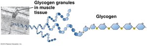
Glycogen: Energy storage in animals (e.g., muscles).
Cellulose: Structural component in plant cell walls.
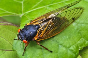
Chitin: Structural component in exoskeletons of insects/crustaceans and fungal cell walls.
Synthesis and Breakdown of Polymers
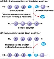
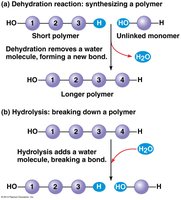
Polymers are synthesized and broken down by specific chemical reactions. Dehydration synthesis forms polymers by removing water, while hydrolysis breaks polymers by adding water.
Dehydration synthesis: Joins monomers, releasing H2O and requiring energy.
Hydrolysis: Breaks polymers, consuming H2O and releasing energy.
Glycosidic Linkages and Disaccharides
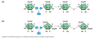
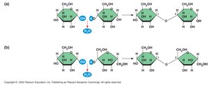
Monosaccharides are covalently joined by glycosidic linkages to form disaccharides through dehydration synthesis. Examples include maltose (glucose + glucose) and sucrose (glucose + fructose).
Starch and Cellulose: Structural Differences
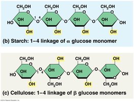
Starch and cellulose are both polymers of glucose but differ in their structure and function due to the type of glucose linkage.
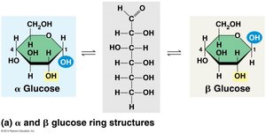
Starch: Composed of α-glucose monomers (energy storage).
Cellulose: Composed of β-glucose monomers (structural support).
Chitin: Structure and Function
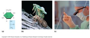
Chitin is a structural polysaccharide similar to cellulose but with an extra functional group. It provides strength to insect exoskeletons and fungal cell walls and is used in medical applications such as surgical threads.
Lipids: Types and Properties
Lipids are hydrophobic molecules with many C-C and C-H bonds, storing energy efficiently. They are not polymers and include triglycerides, phospholipids, and steroids.
Triglycerides: Fats and oils for energy storage.
Phospholipids: Form bilayer membranes.
Steroids: Membrane components and hormones.
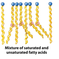
Saturated vs. Unsaturated Fatty Acids
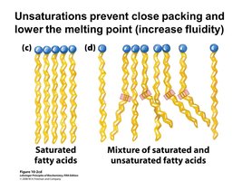
Saturated fats: Pack tightly, solid at room temperature.
Unsaturated fats: Have double bonds, liquid at room temperature.
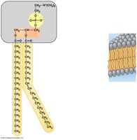
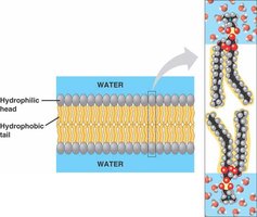
Phospholipids and Membrane Structure
Phospholipids are amphipathic, with hydrophilic heads and hydrophobic tails, forming bilayers that make up cell membranes.
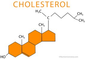
Steroids: Structure and Function
Steroids are organic compounds with four fused rings. Cholesterol is a common steroid in animal cell membranes and a precursor for hormones.
Amino Acids and Proteins
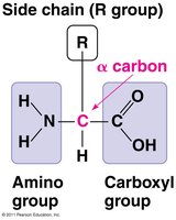
Amino acids are the monomers of proteins, each with a central α-carbon, amino group, carboxyl group, hydrogen, and a unique R-group. There are 20 amino acids classified by their side chains: nonpolar, polar, acidic, and basic.

Protein Structure
Proteins have four levels of structure:
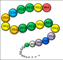
Primary: Sequence of amino acids.
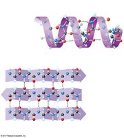
Secondary: Local folding (α-helix, β-sheet) stabilized by hydrogen bonds.
Tertiary: Overall 3D shape due to interactions among R-groups.
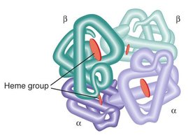
Quaternary: Association of multiple polypeptide chains.
Protein Structure Determination
The sequence of amino acids in proteins is determined by genes encoded in DNA, which directs protein synthesis via messenger RNA (mRNA).
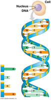
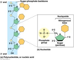
Nucleic Acids: DNA and RNA
Nucleic acids store and transmit genetic information. DNA contains genes for protein expression, and RNA is synthesized from DNA to direct protein synthesis (gene expression).
DNA: Deoxyribonucleic acid, double-stranded, stores genetic information.
RNA: Ribonucleic acid, single-stranded, involved in protein synthesis.
Chapter 6: A Tour of the Cell
Cell Theory and Fundamental Units of Life
All living organisms are composed of cells, which are the basic units of life. Cells arise from pre-existing cells and share common features despite diversity.
Microscopy and Cell Structure
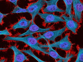
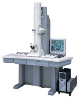
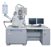
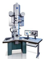
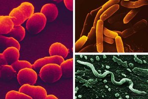
Electron microscopes (EM) are used to view cells at high magnification. Scanning EMs show cell surfaces, while transmission EMs reveal internal structures.
Prokaryotic vs. Eukaryotic Cells
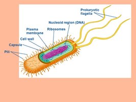
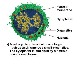
Prokaryotic cells (Bacteria, Archaea) are smaller and simpler, lacking a nucleus and membrane-bound organelles. Eukaryotic cells have a nucleus and organelles.
Membrane-Bound Organelles
Eukaryotic cells contain organelles such as the nucleus, ribosomes, endoplasmic reticulum, Golgi apparatus, lysosomes, vacuoles, and peroxisomes, each with specialized functions.
Nucleus and Ribosomes
Nucleus: Contains DNA, controls cell activities, directs protein synthesis.
Nucleolus: Site of ribosomal RNA synthesis.
Ribosomes: Make proteins, found free in cytosol or bound to ER.
Endoplasmic Reticulum (ER)
Rough ER: Synthesizes proteins for export.
Smooth ER: Synthesizes lipids, detoxifies substances, stores calcium.
Golgi Apparatus
Modifies and packages proteins from ER.
Receives and ships vesicles.
Lysosomes and Vacuoles
Lysosomes: Digestive compartments, break down food, bacteria, and old organelles.
Vacuoles: Storage and osmoregulation in protists and plants.
Peroxisomes
Break down fatty acids, detoxify harmful substances.
Energy Processing Organelles
Mitochondria: Power plant of cell, site of cellular respiration.
Chloroplasts: Site of photosynthesis in plants.
Endomembrane System
The endomembrane system includes the nuclear envelope, ER, Golgi apparatus, lysosomes, vacuoles, and plasma membrane, connected directly or via vesicles.
Cytoskeleton
The cytoskeleton provides structural support, movement, and communication. It consists of microtubules, intermediate filaments, and microfilaments.
Cell Communication and Junctions
Tight junctions: Prevent leakage of fluid.
Anchoring junctions: Fasten cells together.
Gap junctions: Allow small molecules to flow between cells.
Chapter 7: Membrane Structure and Function
Plasma Membrane and Selective Permeability
The plasma membrane separates the cell from its environment and regulates the passage of substances. It is composed of a fluid mosaic of phospholipids and proteins.
Membrane Fluidity
Membrane fluidity is influenced by the composition of phospholipids and cholesterol. Unsaturated fatty acids increase fluidity by preventing tight packing.
Transport Across Membranes
Molecules cross membranes by passive or active transport.
Passive transport: Diffusion of small molecules (e.g., O2, CO2) down concentration gradients, no energy required.
Active transport: Movement against concentration gradient, requires energy (ATP).
Diffusion and Dynamic Equilibrium
Diffusion is the movement of molecules from high to low concentration until dynamic equilibrium is reached.
Water Transport and Membrane Selectivity
Water moves across membranes via aquaporins, specialized transport proteins. Membrane selectivity ensures proper water balance.
Tonicity and Water Balance
Tonicity describes the effect of solute concentration on water movement:
Isotonic: Equal solute concentration inside and outside cell.
Hypotonic: Lower solute outside; water enters cell.
Hypertonic: Higher solute outside; water leaves cell.
Active Transport and ATP
Active transport uses ATP to move solutes against their concentration gradient, maintaining essential cellular functions.
Additional info: All explanations are expanded for academic completeness and clarity. Images are included only when directly relevant to the adjacent content.
