 Back
Study Guide: The Molecular Basis of Inheritance – DNA Structure and Replication
Study Guide - Smart Notes
 Tailored notes based on your materials, expanded with key definitions, examples, and context.
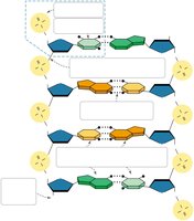
Q8. Label the diagram of the DNA double helix, noting the antiparallel structure and chemical details.
Background
Topic: DNA Structure
This question tests your understanding of the molecular structure of DNA, including the antiparallel arrangement of the two strands, the sugar-phosphate backbone, base pairing, and the types of bonds involved.
Key Terms and Concepts:
Antiparallel: The two strands of DNA run in opposite directions (5' to 3' and 3' to 5').
Sugar-phosphate backbone: The repeating chain of deoxyribose sugars and phosphate groups.
Base pairs: Adenine (A) pairs with Thymine (T), and Guanine (G) pairs with Cytosine (C) via hydrogen bonds.
Phosphodiester bonds: Covalent bonds linking the sugar and phosphate groups in the backbone.
Hydrogen bonds: Weak bonds between complementary nitrogenous bases.
Step-by-Step Guidance
Identify the sugar-phosphate backbone on each strand. These are the outermost chains, composed of alternating deoxyribose sugars and phosphate groups.
Locate the nitrogenous bases (A, T, G, C) that extend inward from the backbone. Each base on one strand pairs with a complementary base on the opposite strand.
Note the directionality of each strand: one runs from 5' to 3', and the other from 3' to 5'. This antiparallel orientation is crucial for DNA function.
Label the hydrogen bonds (dotted lines) between complementary bases: A pairs with T (2 hydrogen bonds), G pairs with C (3 hydrogen bonds).
Indicate the phosphodiester bonds (solid lines) connecting the sugar and phosphate groups along each backbone.
Try solving on your own before revealing the answer!
Q12. What does it mean that the two strands of DNA are antiparallel?
Background
Topic: DNA Structure – Antiparallel Orientation
This question focuses on the orientation of the two DNA strands and why this arrangement is important for replication and function.
Key Terms:
Antiparallel: Refers to the opposite orientation of the two DNA strands.
5' end: The end of the DNA strand with a free phosphate group attached to the 5' carbon of the sugar.
3' end: The end with a free hydroxyl group attached to the 3' carbon of the sugar.
Step-by-Step Guidance
Recall that each DNA strand has a directionality, determined by the carbon atoms in the deoxyribose sugar (numbered 1' to 5').
One strand runs from the 5' end (phosphate group) to the 3' end (hydroxyl group), while the complementary strand runs in the opposite direction (3' to 5').
This antiparallel arrangement allows the bases to pair correctly and is essential for the enzymes involved in DNA replication and repair to function properly.
Try solving on your own before revealing the answer!
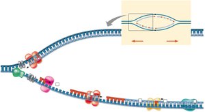
Q21. Label the DNA replication fork diagram with the following: 3' and 5' strands, RNA primer, primase, SSBP, topoisomerase, helicase, leading strand, lagging strand, DNA pol I, DNA pol III, DNA ligase, parental DNA, and new DNA. Add notes explaining replication on both strands.
Background
Topic: DNA Replication
This question tests your ability to identify the key enzymes and structures involved in DNA replication and to understand the process on both the leading and lagging strands.
Key Terms and Concepts:
Replication fork: The Y-shaped region where the DNA is split into two strands for copying.
Leading strand: Synthesized continuously in the 5' to 3' direction.
Lagging strand: Synthesized discontinuously as Okazaki fragments.
Key enzymes: Helicase, primase, DNA polymerase III, DNA polymerase I, DNA ligase, topoisomerase, single-strand binding proteins (SSBP).
Step-by-Step Guidance
Identify the parental DNA strands and the directionality (3' and 5' ends) of each strand at the replication fork.
Locate the leading strand (synthesized continuously) and the lagging strand (synthesized in fragments).
Label the enzymes: helicase (unwinds DNA), primase (synthesizes RNA primer), DNA polymerase III (extends new DNA), DNA polymerase I (replaces RNA primer with DNA), DNA ligase (joins fragments), topoisomerase (relieves strain), and SSBP (stabilizes single strands).
Indicate the RNA primers and Okazaki fragments on the lagging strand.
Add notes explaining that the leading strand is synthesized continuously toward the replication fork, while the lagging strand is synthesized away from the fork in short fragments.
Try solving on your own before revealing the answer!
