 Back
BackThe Cell Cycle: Mechanisms, Regulation, and Cancer
Study Guide - Smart Notes
 Tailored notes based on your materials, expanded with key definitions, examples, and context.
The Cell Cycle
Introduction to Cell Division
Cell division is a fundamental process by which a single parent cell divides to produce two or more daughter cells. This process is essential for reproduction, growth, and tissue repair in all living organisms. There are three main types of cell division:
Binary Fission: A form of asexual cell division in prokaryotes (bacteria and archaea).
Mitosis: Eukaryotic cell division that produces genetically identical somatic (body) cells.
Meiosis: Eukaryotic cell division that produces genetically unique gametes (sex cells).
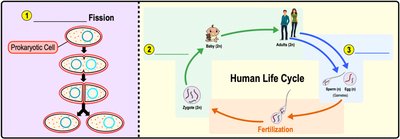
Example: The human life cycle involves both mitosis (for growth and repair) and meiosis (for gamete production).
Asexual vs. Sexual Reproduction
Organisms reproduce either asexually or sexually, each with distinct genetic outcomes:
Asexual Reproduction: Involves one parent and produces genetically identical offspring (clones). Common in prokaryotes and some eukaryotes.
Sexual Reproduction: Involves two parents and produces genetically diverse offspring due to the combination of genetic material from both parents.
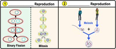
Example: Binary fission in bacteria is asexual, while meiosis and fertilization in humans are sexual.
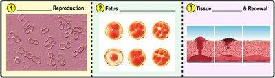
Importance of Cell Division
Cell division is crucial for:
Reproduction: Producing new organisms.
Growth and Development: Increasing cell number during development.
Tissue Repair and Renewal: Replacing damaged or dead cells.
Before division, a cell must replicate its DNA to ensure each daughter cell receives a complete set of genetic instructions.
Organization of DNA in the Cell
Genome Structure
The genome is the complete set of an organism's DNA. In eukaryotes, DNA is organized with proteins called histones into units called nucleosomes. The structure of DNA changes depending on the cell's stage:
Chromatin: Loosely packed DNA in non-dividing cells.
Chromosomes: Highly condensed DNA in dividing cells.
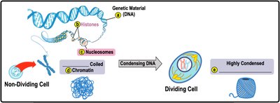
Example: Chromatin condenses into visible chromosomes during cell division.
DNA Replication and Chromosome Structure
Before a cell divides, it replicates its DNA, resulting in chromosomes composed of two identical sister chromatids joined at a centromere.
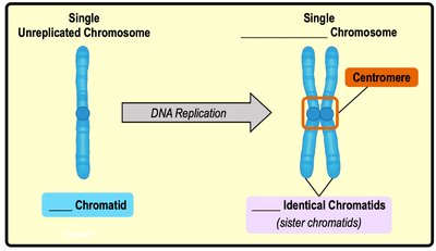
Chromatid: One of two identical halves of a replicated chromosome.
Sister Chromatids: Genetically identical copies joined together until separated during cell division.
The Cell Cycle: Phases and Regulation
Phases of the Cell Cycle
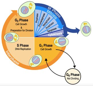
The cell cycle describes the sequence of events from the formation of a cell to its division into daughter cells. It consists of two major phases:
Interphase: Non-dividing phase for cell growth, DNA replication, and preparation for division. Subdivided into G1, S, and G2 phases.
M Phase (Mitotic Phase): Dividing phase, including mitosis (nuclear division) and cytokinesis (cytoplasmic division).
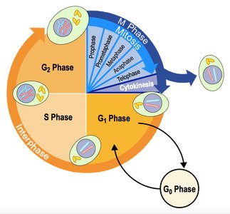
Sub-phases of Interphase
Interphase is divided into:
G1 Phase (First Gap): Cell growth and normal function.
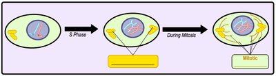
S Phase (Synthesis): DNA replication and centrosome duplication.
G2 Phase (Second Gap): Further growth and preparation for mitosis.
G0 Phase: Non-dividing state for cells that have exited the cycle.
Centrosomes and Mitotic Spindle
During S phase, the centrosome is duplicated. The centrosome organizes the mitotic spindle, a structure made of microtubules that separates chromosomes during mitosis.
Mitosis: Phases and Mechanisms
Overview of Mitosis
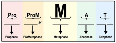
Mitosis is the process of dividing the nucleus and genetic material of a somatic cell, resulting in two genetically identical daughter cells. Mitosis consists of five phases:
Prophase
Prometaphase
Metaphase
Anaphase
Telophase
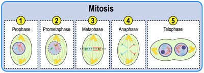
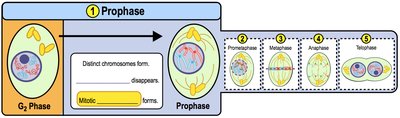
Prophase
During prophase, chromatin condenses into visible chromosomes, the nucleolus disappears, and centrosomes move to opposite poles, beginning spindle formation.
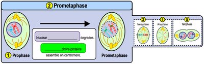
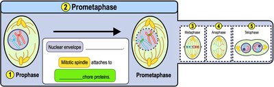
Prometaphase
The nuclear envelope breaks down, exposing chromosomes to the cytoplasm. Spindle fibers attach to chromosomes at the kinetochore, a protein complex at the centromere.
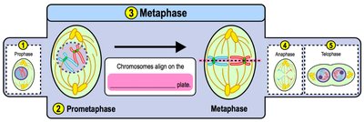
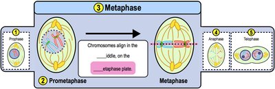
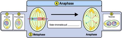
Metaphase
Chromosomes align at the metaphase plate (cell equator), with spindle fibers attached to each sister chromatid.
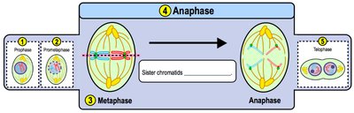
Anaphase
Sister chromatids are separated and pulled toward opposite poles of the cell by shortening spindle fibers. Non-kinetochore microtubules elongate the cell.
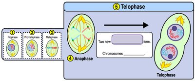
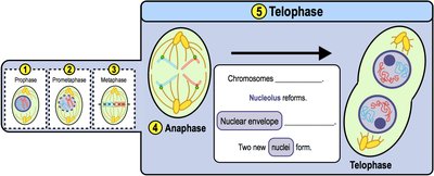
Telophase
Telophase is the reverse of prophase: chromosomes decondense, the nuclear envelope and nucleolus reform, and two nuclei are established in the cell.
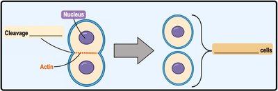
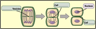
Cytokinesis
Animal Cell Cytokinesis
In animal cells, cytokinesis occurs via the formation of a cleavage furrow, where actin and myosin filaments contract to pinch the cell into two daughter cells.
Plant Cell Cytokinesis
In plant cells, vesicles from the Golgi apparatus coalesce at the center of the cell to form a cell plate, which develops into a new cell wall, separating the two daughter cells.
Regulation of the Cell Cycle
Cell Cycle Checkpoints
Cell division is tightly regulated by growth factors and cell cycle checkpoints that ensure each phase is completed correctly before the next begins. The main checkpoints are:
G1 Checkpoint: Checks for DNA damage before replication.
G2 Checkpoint: Ensures DNA replication is complete and correct.
M (Metaphase) Checkpoint: Confirms all chromosomes are properly attached to the spindle before separation.
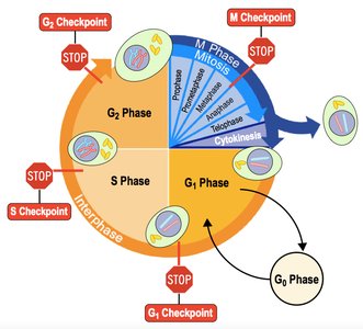
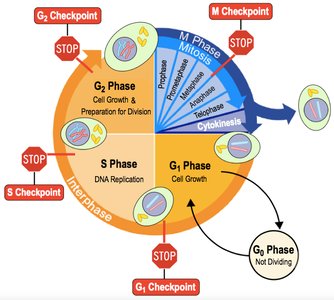
If errors are detected, proteins like p53 can trigger repair or apoptosis (programmed cell death). Failure in checkpoint control can lead to uncontrolled cell division and cancer.
Cancer and the Cell Cycle
Uncontrolled Cell Division and Tumors
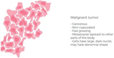
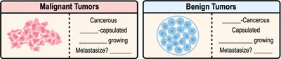
Cancer is characterized by uncontrolled cell division, leading to the formation of tumors. Tumors can be:
Benign: Non-cancerous, localized, and slow-growing.
Malignant: Cancerous, invasive, fast-growing, and capable of metastasis (spreading to other tissues).
Genes Regulating Cell Growth
Two main types of genes regulate cell growth:
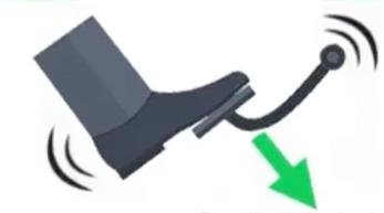
Proto-oncogenes: Promote normal cell division ("gas pedal"). Mutations can convert them into oncogenes, which drive uncontrolled division.
Tumor Suppressor Genes: Inhibit cell division ("brakes"). Mutations can disable these genes, removing growth inhibition.
Example: The p53 gene is a well-known tumor suppressor gene.
Summary Table: Benign vs. Malignant Tumors
Feature | Benign Tumor | Malignant Tumor |
|---|---|---|
Cancerous? | No | Yes |
Capsulated? | Yes | No |
Growth Rate | Slow | Fast |
Metastasis? | No | Yes |
Cell Appearance | Normal | Abnormal, large nuclei |
Additional info: Mutations in proto-oncogenes or tumor suppressor genes can disrupt the balance of cell division, leading to cancer. Proper regulation of the cell cycle is essential for organismal health and prevention of disease.
