 Back
The Cell Cycle: Mechanisms, Regulation, and Clinical Relevance chpt.12
Study Guide - Smart Notes
 Tailored notes based on your materials, expanded with key definitions, examples, and context.
The Cell Cycle and Cell Division
Overview of Cell Division
The cell cycle is the series of events that cells go through as they grow and divide. Cell division is essential for reproduction, growth, development, and tissue renewal in multicellular organisms.
Reproduction: Unicellular organisms reproduce by dividing into two new cells.
Growth and Development: Multicellular organisms grow from a single cell to trillions of cells through repeated cell divisions.
Tissue Renewal: Cells divide to replace old, damaged, or dead cells, maintaining tissue health.
The Cell Cycle Phases
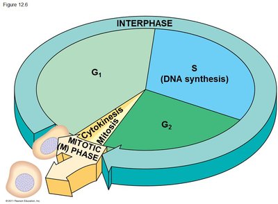
The cell cycle consists of a sequence of phases: Interphase (G1, S, G2) and the Mitotic (M) phase, which includes mitosis and cytokinesis.
G1 phase (First Gap): Cell grows and carries out normal functions.
S phase (Synthesis): DNA is replicated, resulting in two identical sets of chromosomes.
G2 phase (Second Gap): Cell prepares for mitosis by producing proteins and organelles; centrosomes duplicate.
M phase (Mitosis and Cytokinesis): The cell divides its nucleus and cytoplasm to form two genetically identical daughter cells.
Genetic Material and Chromosome Structure
Chromatin and Chromosomes
Genetic information is stored in DNA, which is packaged with proteins to form chromatin. During cell division, chromatin condenses into visible chromosomes.
Chromatin: DNA-protein complex that is loosely packed during interphase.
Chromosome: Densely packed chromatin, visible during mitosis.
Histones: Proteins that help organize DNA into nucleosomes.
Chromosome Number and Types
Each species has a characteristic number of chromosomes. In humans, somatic cells have 46 chromosomes (23 pairs), while gametes have 23 chromosomes.
Somatic Cells: Body cells with pairs of homologous chromosomes (one from each parent).
Gametes: Sex cells (sperm and eggs) with one chromosome from each pair.
Homologous Chromosomes: Chromosome pairs with the same genes but possibly different alleles.
DNA Replication and Sister Chromatids
Before a cell divides, it must replicate its DNA so that each daughter cell receives an identical set of chromosomes. Each replicated chromosome consists of two sister chromatids joined at the centromere.
Sister Chromatids: Identical copies of a chromosome, attached at the centromere.
Centromere: Region where sister chromatids are most closely attached.
Phases of Mitosis
Stages of Mitosis
Mitosis is the process by which a eukaryotic cell separates its duplicated chromosomes into two identical nuclei. It consists of several stages:
Prophase: Chromatin condenses into chromosomes; mitotic spindle begins to form; nucleoli disappear.
Prometaphase: Nuclear envelope breaks down; spindle microtubules attach to kinetochores on chromosomes.
Metaphase: Chromosomes align at the metaphase plate; spindle fibers attach to kinetochores from opposite poles.
Anaphase: Sister chromatids separate and move toward opposite poles; cell elongates.
Telophase: Nuclear envelopes reform; chromosomes decondense; nucleoli reappear.
Cytokinesis: Division of the cytoplasm, resulting in two daughter cells.
Cytokinesis
Cytokinesis is the division of the cytoplasm, which follows mitosis and results in two separate daughter cells.
Animal Cells: Form a cleavage furrow, which pinches the cell in two using a contractile ring of actin microfilaments.
Plant Cells: Form a cell plate from vesicles that fuse at the center, eventually developing into a new cell wall.
The Mitotic Spindle and Chromosome Movement
Mitotic Spindle Structure and Function
The mitotic spindle is a structure made of microtubules that segregates chromosomes during mitosis.
Centrosome: Microtubule-organizing center; duplicates during G2 phase.
Asters: Radial arrays of short microtubules extending from centrosomes.
Kinetochore Microtubules: Attach to kinetochores on chromosomes and pull sister chromatids apart.
Nonkinetochore Microtubules: Overlap at the cell center and help elongate the cell during anaphase.
Kinetochore Structure
The kinetochore is a protein complex assembled on the centromere of each chromosome, serving as the attachment site for spindle microtubules.
Inner and Outer Kinetochore: Layers of proteins that interact with centromeric DNA and microtubules.
Function: Ensures proper chromosome segregation by attaching chromosomes to spindle fibers.
Regulation of the Cell Cycle
Cell Cycle Control System
The cell cycle is regulated by a complex control system involving checkpoints, cyclins, and cyclin-dependent kinases (Cdks).
Checkpoints: Control points where the cell cycle can be stopped until conditions are favorable (G1, G2, and M checkpoints).
Cyclins: Regulatory proteins whose levels fluctuate during the cell cycle.
Cyclin-Dependent Kinases (Cdks): Enzymes activated by cyclins; phosphorylate target proteins to advance the cell cycle.
MPF (Maturation-Promoting Factor): A cyclin-Cdk complex that triggers entry into mitosis.
Key Equations:
MPF activity is regulated by cyclin concentration:
Major Cell Cycle Checkpoints
G1 Checkpoint (Restriction Point): Determines if the cell will divide; if not, the cell enters G0 (non-dividing state).
G2 Checkpoint: Ensures DNA replication is complete and undamaged before mitosis.
M Checkpoint: Ensures all chromosomes are properly attached to the spindle before anaphase begins.
External Signals and Cell Cycle Regulation
Growth Factors: Proteins such as PDGF stimulate cell division in certain cell types.
Density-Dependent Inhibition: Cells stop dividing when they contact neighboring cells.
Anchorage Dependence: Cells must be attached to a substrate to divide.
Cancer and the Cell Cycle
Loss of Cell Cycle Control in Cancer
Cancer cells escape normal cell cycle controls, leading to uncontrolled proliferation.
Characteristics: Ignore growth factors, do not respond to stop signals, avoid apoptosis, and can divide indefinitely (immortal cell lines, e.g., HeLa cells).
Tumors: Benign tumors remain localized; malignant tumors invade other tissues (metastasis).
Genetic Changes: Cancer cells often have abnormal chromosome numbers and altered metabolism.
Cancer Treatment and Prevention
Radiation: Damages DNA in rapidly dividing cells.
Chemotherapy: Targets dividing cells throughout the body.
Personalized Medicine: Uses genetic information to tailor treatments (e.g., Cancer Genome Project).
Prevention: Avoiding carcinogens such as tobacco smoke and excessive UV exposure reduces cancer risk.
Summary Table: Key Terms and Concepts
Term | Definition |
|---|---|
Chromatin | DNA-protein complex in the nucleus |
Chromosome | Condensed chromatin visible during cell division |
Sister Chromatids | Identical copies of a chromosome joined at the centromere |
Centromere | Region where sister chromatids are attached |
Mitotic Spindle | Microtubule structure that separates chromosomes |
Cytokinesis | Division of the cytoplasm |
Checkpoint | Control point in the cell cycle |
Cyclin | Regulatory protein controlling cell cycle progression |
Cdk | Enzyme activated by cyclin to regulate the cell cycle |
MPF | Cyclin-Cdk complex that triggers mitosis |
Benign Tumor | Non-invasive, localized tumor |
Malignant Tumor | Invasive tumor that can spread (cancer) |
Practice Questions
The drug cytochalasin B blocks the function of actin. Which aspect of the animal cell cycle would be most disrupted by cytochalasin B? Answer: E. Cleavage furrow formation and cytokinesis
Which of the following does not occur during mitosis? Answer: A. Replication of the DNA
The decline of MPF activity at the end of mitosis is due to Answer: B. The degradation of cyclin
